Index and Table of Contents
Technical and Practice Issues
- Case 11: 62-years old misdiagnosed to have interstitial lung disease - mid-inspiratory and expiratory scans
- Snippet 03: Radiation risk and CT chest
- Case 23: 60-years old - 40-pack years smoker - right upper lobe nodule - resolved using "mean" reconstructions to simulate a chest radiograph
- Snippet 11: ACR appropriateness criteria for diffuse lung disease imaging and ACR-STR guidelines
- Snippet 22: What defines an ideal CT scan for diffuse lung diseases
- Dual energy CT (DECT)
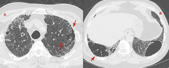
- Case 14: 70-years old man with COVID-19 - DECT showing perfusion defects
- Case 19: 55-years old with COVID-19 vasculopathy / angiopathy - 10 1/2 months follow-up
- Case 24: 55-years old - perfusion defects in chronic thromboembolic pulmonary hypertension (CTEPH)
- Case 38: 40-years old man with COVID-19 - 15 months follow-up of COVID changes and vasculopathy
- Case 102: 76-years old with tracheobronchial tuberculosis - the uses of minimum intensity projection (minIP) reconstructions
- Case of the Day 096: 56-years old suspected to have ILD because of "ground glass" diagnosed on an expiratory scan - pitfalls of a poor quality scan
- Case of the Day 162: 70 years old with interstitial lesions. Real or mimics
- Case of the Day 165: 60-yers old lady with ILD and a left axillary node - metastatic breast carcinoma - all about looking for breast lesions on CT chest
Principles of Interpretation
(Principles / Approach / Signs / Indications)
-
Signs and Differentials
- Snippet 01: Differentiating traction bronchiectasis from honeycombing
- Snippet 02: Centrilobular / bronchocentric nodules
- Snippet 04: Fibrotic NSIP vs probable UIP
- Case 16: 56-years old - Approach to interpretation of diffuse ground glass
- Case 17: 17-years old with hemoptysis - tuberculosis - differentiating dense centrilobular nodules of infectious bronchiolitis from soft nodules due to blood
- Snippet 06: Six fibrosing ILD questions answered
- Snippet 07: CT signs suggesting connective tissue disease (CTD) ILD
- Snippet 08: Mosaic attenuation - differentiating CTEPH, small airways disease and interstitial lung disease
- Snippet 09: Variant fibrosis in connective tissue disease (CTD) ILD
- Snippet 10: Diagnosis of bronchiectastis
- Snippet 12: Visual classification of centrilobular and paraseptal emphysema
- Snippet 13: Spectrum of smoking related changes in a single patient
- Case 66: Importance of the inspiratory-expiratory pair
- Case 68: Looking at the Lungs Carefully When There is Lymphadenopathy
- Snippet 15: CT Scan Findings in Sarcoidosis
- Lecture: Signs of Fibrosing ILDs - Reticular opacities, traction bronchiectasis and honeycombing
- Lecture: Approach to diffuse small lung nodules
- Lecture: Overview of diffuse lung diseases
- Lecture: Centrilobular nodule, proliferative bronchiolitis, etc
- Snippet 16: Differentiating inflammatory from fibrotic ILDs
- Snippet 17: How to diagnose subtle progression of interstitial lung disease at the secondary pulmonary lobule level
- Case 91: Bubble lucencies, central lucencies, bubbly consolidation - causes and differentials
- Snippet 18: The jellyfish and multiple pleural tag signs to predict visceral pleural invasion in small lung cancers
- Snippet 20: Practical approach to diffuse cystic lung diseases
- Snippet 21: Approach to right medial cardiophrenic angle lesions
- Case of the Day 165: 60-yers old lady with ILD and a left axillary node - metastatic breast carcinoma - all about looking for breast lesions on CT chest
-
First Principles of Interpretation
- Do not ignore the non-contrast (plain) scan - 3 cases
- Case 33: 70-years old - looking at the plain scan to resolve active nodes on PET/CT - old healed granulomatous infection
- Case 50: First princples - look at the noncontrast (plain) scan subcarinal bronchogenic cyst
- Case 55: First principles - trace the bronchus - non-resolving consolidation - foreign body - clove in left lower lobe bronchial segment
- Case 69: What ails chest imaging training? 60-years old with a left upper lobe lesion for biopsy
- Case 107: Why LLMs will render many radiologists and their reports irrelevant if they don't up their game - UIP/IPF
- Case 108: Approaching a patient with PH - it can't be so difficult - PAPVR and Gr 2 PH
- Case of the Day 093: Why AI is the Only Solution to Our Chest X-ray Hyposkillia
-
Indications
Guidelines and White Papers and Oration Lectures
- 2020 ATS/JRS/ALAT - Hypersensitivity pneumonitis - new classification
- Snippet 11: ACR appropriateness criteria for diffuse lung disease imaging and ACR-STR guidelines
- 2022 ATS/ERS/JRS/ALAT - Progressive pulmonary fibrosis - fibrotic hypersensitivity pneumonitis
- 2022 ATS/ERS/JRS/ALAT - Progressive pulmonary fibrosis - scleroderma ILD
- 2022 ATS/ERS/JRS/ALAT - CPFE - Official statement
- Lecture: Dr S K Bhalla Oration at SCIICON 2024 - Evolution of HRCT - A 33-Years Journey
- 2025 ATS clinical position statement on interstitial lung abnormalities (ILAs)
Lung
-
Interstitial Lung Diseases
-
ILD Patterns (A New Series from Dec 2025)
-
The UIP Pattern - 28 Dec 2025
-
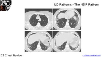
The NSIP Pattern - 24 Mar 2026
-
The Variant UIP Pattern - 17 May 2026
-
Fibrotic
-
- UIP/IPF
- Case 5: 70-years old male smoker - usual interstitial pneumonia (UIP) / idiopathic pulmonary fibrosis (IPF)
- Case 6: 63-years old man - probable usual interstitial pneumonia (probable UIP) / idiopathic pulmonary fibrosis (IPF)
- Case 7: 75-years old man - UIP/IPF with a complication - lung nodule - squamous cell carcinoma
- Snippet 01: Differentiating traction bronchiectasis from honeycombing
- Snippet 04: Fibrotic NSIP vs probable UIP
- Snippet 06: Six fibrosing ILD questions answered
- Case 90: 57-years old - probable UIP/IPF - progressive disease - how time matters
- Case 92: 67-years old - probable UIP/IPF - acute exacerbation, likely triggered
- Case 98: 73-years old - UIP/IPF with acute increased dyspnea due to pulmonary edema and cardiac failure
- Case 107: 76-years old - UIP/IPF - why LLMs will render many radiologists and their reports irrelevant
- Live Case 04 - Feb 2026: 65-years old with UIP-IPF
- Familial Pulmonary Fibrosis (FPF)
- Combined Pulmonary Fibrosis and Emphysema (CPFE)
- Snippet 13: Spectrum of smoking related changes in a single patient
- Case 48: 78-years old - combined pulmonary fibrosis and emphysema (CPFE)
- Case 67: 69-years old - combined pulmonary fibrosis and emphysema (CPFE) and the official statement from ATS/ERS/JRS/ALAT
- Snippet 27: Airspace Enlargement with Fibrosis (AEF)
- Fibrotic NSIP / Variant Fibrosis / Connective Tissue Disease ILD
- Snippet 04: Fibrotic NSIP vs probable UIP
- Case 10: 67-years old woman - fibrotic organizing pneumonia (OP) - Sjogren's
- Snippet 07: CT signs suggesting connective tissue disease (CTD) ILD
- Snippet 09: Variant fibrosis in connective tissue disease (CTD) ILD
- Case 39: 27-years old - scleroderma ILD - secondary pleuroparenchymal fibroelastosis (PPFE)
- Case 58: 27-years old - scleroderma ILD - progressive pulmonary fibrosis
- Case 94: 24-years old - scleroderma ILD - early/subtle disease
- Lecture: NSIP and OP
- Lecture: Connective tissue disease ILDs
- Case 111: 59-years old woman - lymphoid interstitial pneumonia (LIP) - Sjogren's
- Case of the Day 066: 60-years old - rheumatoid arthritis (RA) ILD with a usual interstitial pneumonia (UIP) pattern with variant fibrosis with superimposed CCPA
- Case of the Day 093: Why AI is the only solution to our chest X-ray hyposkillia - Rheumatoid arthritis-Sjogren ILD with a variant UIP pattern with superimposed CCPA as a complication
- Snippet 28: Connective tissue disease ILD patterns
- Case of the Day 116: 31-yrs old with scleroderma and a mixed BIP/NSIP/CiOP pattern - mixed patterns in connective tissue disease ILDs
- Case of the 117: 51-yrs old in 2012 with a mixed progressive fibrotic NSIP/OP/DPO patter, diagnosed to have scleroderma/MCTD 13 years later
- Case of the Day 123: 46-yrs old with anti-synthetase syndrome and a progressive NSIP pattern showing the peri-diaphragmatic inflammation & fibrosis - PDIF sign
- Post 01: 59-years old with indeterminate fibrosing ILD but with likely immune-dysregulation related ILD with new diffuse ground glass
- Live Case 05 - Feb 2026: 63-years old woman with scleroderma ILD with an NSIP pattern
- Post 04: 64-years old smoker with RA - RA, RA-ILD, AEF and CPFE
- Case of the Day 174: 33-years old with scleroderma showing an NSIP pattern along with FANO - faint amorphous nodular opacification
- Non-UIP/Non-IPF/Bronchiolocentric Interstitial Pneumonia (BIP)
- Fibrotic Hypersensitivity Pneumonitis
- Case 49: 65-years old - fibrotic hypersensitivity pneumonitis - hexagonal pattern
- Case 57: 55-years old man - fibrotic hypersensitivity pneumonitis - progressive pulmonary fibrosis
- Case 85: 65-years old non-smoker - fibrotic hypersensitivity pneumonitis
- Lecture 08: CT in hypersensitivity pneumonitis
- Case of the Day 086: 56-years old with fibrotic hypersensitivity pneumonitis - discussion of three density sign and the new label of the bronchiolocentric interstitial pneumonia (BIP) pattern
- P*ost 02:70-years old with a progressive bronchiolocentric interstitial pneumonia (BIP) pattern due to fibrotic hypersensitivity pneumonitis over 10 years - progressive pulmonary fibrosis*
- Chronic Pulmonary Sarcoidosis
- Fibrotic Hypersensitivity Pneumonitis
- Indeterminate
- Miscellaneous
- UIP/IPF
-
Nonfibrotic
- Case 16: 56-years old - PCP / PJP in a PLHA presenting as diffuse ground glass
- Case 26: 16-years old - pulmonary alveolar proteinosis (PAP)
- Case 46: 70-years old - nitrofurantoin lung injury with organizing pneumonia (OP) pattern
- Case 59: 73-years old - nivolumab lung injury - pre-existing ILA
- Case 85: 65-years old non-smoker - non-fibrotic hypersensitivity pneumonitis progressing to fibrotic
- Lecture: REF-Cafe Roentgen Lecture 03 - Non-fibrosing ILDs
- Lecture 08: CT in hypersensitivity pneumonitis
- Lecture: NSIP and OP
- Case of the Day 092: 44-years old - PCP/PJP in a PLHA presenting as just non-specific ground glass along with non-pulmonary findings
- Case 115: 36-years old - ground glass plus septal thickening - lymphangitis from signet call adenocarcinoma
- Case of the Day 131: 48-yrs old with classic crazy paving - pulmonary alveolar proteinosis
-
ARDS/AIP/Acute Lung Injury/Diffuse Alveolar Hemorrhage/Acute Diffuse Infection
- Case 44: 33-years old - ANCA-associated vasculitis and diffuse alveolar hemorrhage
- Case 64: 70-years old - H1N1 lung involvement - organizing pneumonia (OP) pattern
- Case 92: 67-years old - probable UIP/IPF - acute exacerbation, likely triggered
- Case 97: 38-year old - miliary tuberculosis presenting as ARDS
- Case of the Day 074: 57-yrs old - acute lung injury - exudative to organizing phase
- Post 01: 59-years old with indeterminate fibrosing ILD but with likely immune-dysregulation related ILD with new diffuse ground glass
-
Interstitial Lung Abnormality (ILA)
- Case 36: 76-years old - interstitial lung abnormality (ILA) to fibrotic interstitial lung disease (ILD)
- Case 59: 73-years old - nivolumab lung injury - pre-existing ILA
- Lecture 10: How to approach a patient with interstitial lung abnormality - ILA
- Snippet 25: Interstitial lung abnormality (ILA) - what is new and what has changed
- COD 118: 68-yrs old with indeterminate interstitial lesions and previous COVID-19 exposure simulating an ILA
- Lecture 16: Interstitial lung abnormality (ILA) update
-
Nodular
- Case 12: 38-years old - nodules and septal thickening - acute onset - tropical pulmonary eosinophilia
- Snippet 02: Centrilobular / bronchocentric nodules
- Case 29: 58-years old - sarcoidosis over 18 years
- Case 30: 51-years old - classic silicosis
- Case 68: 47-years old - Stage II sarcoidosis
- Lecture: Approach to diffuse small lung nodules
- Case 84: 30-years old - classic miliary tuberculosis
- Case 88: 48-years old - flour mill worker silicosis
- Case 97: 38-year old - miliary tuberculosis presenting as ARDS
- Case 101 - 35-years old - miliary tuberculosis coexisting with infectious bronchiolitis and tree-in-bud lesions - disseminated tuberculosis
- Case 105: 4 different patients - pseudo-miliary appearance - tropical pulmonary eosinophilia
- Case of the Day 172: 49-years old - perivascular, fissural and subpleural nodules with adenopathy - classic stage II sarcoidosis
- Case of the Day 176: 65-years old - pembrolizumab lung injury with organizing pneumonia pattern showing the atoll sign - reversed ground glass
- Case of the Day 187: 71 years old with miliary tuberculosis with a large broncholith in the right main bronchus
- Post 09: 60-years old flour-mill worker with variable-sized small nodules - silicosis, not miliary tuberculosis
-
Miscellaneous
- Case 3: 19-years old man with lungs of stone - alveolar microlithiasis
- Case 4: 70-years old with carcinoma breast - lymphangitis carcinomatosis
- Case 60: 69-years old - radiation pneumonitis and fibrosis
- Case 70: 51-years old - idiopathic diffuse pulmonary ossification (DPO)
- Case 79: 74-years old man - radiation recall pneumonitis
- Case 116: 63-years old - idiopathic dendriform diffuse pulmonary ossification (DPO)
- Snippet 29: IgG4-related disease
-
-
Cystic Lung Diseases
- Case 35: 46-years old - cysts and nodules - amyloidosis
- Case 65 - 46-years old - Birt-Hogg-Dube (BHD) syndrome
- Lecture: REF-Cafe Roentgen Lecture 07 - Diffuse cystic lung diseases
- Snippet 20: Practical approach to diffuse cystic lung diseases
- Case of the Day 083: 31-years old - cysts and lymphangiomas - lymphangioleiomyomatosis - LAM
-
Childhood ILDs (chILD)
-
Emphysema, COPD, Asthma
-
Emphysema/COPD
- Snippet 12: Visual classification of centrilobular and paraseptal emphysema
- Snippet 13: Spectrum of smoking related changes in a single patient
- Case 48: 78-years old - combined pulmonary fibrosis and emphysema (CPFE)
- Case 54: 56-years old - never-smoker - COPD - severe small airways disease
- Case 67: 69-years old - combined pulmonary fibrosis and emphysema (CPFE) and the official statement from ATS/ERS/JRS/ALAT
- Case of the Day 073: 74-years old - never-smoker with COPD, centrilobular emphysema, AEF and CPFE
- Snippet 27: Airspace Enlargement with Fibrosis (AEF)
-
Asthma
-
-
Focal Lung Diseases
-
Solitary Nodule
- Case 1C: 67-years old with a calcified lung nodule - old granuloma - part of the "do not ignore the plain scan" series
- High risk CT imaging features in lung cancer screening - lecture
- Case 7: 75-years old man - UIP/IPF with a complication - lung nodule - squamous cell carcinoma
- Case 23: 60-years old - 40-pack years smoker - right upper lobe nodule - old granuloma
- Case 31: 54-years old - incidental lung nodule - pulmonary sclerosing pneumocytoma
- Case 52: 54-years old - solitary nodule with cyst - neuroendocrine tumor - carcinoid
- Case 76: 57-years old - solitary perifissural nodule (PFN)
- Case 86: 55-years old - growing cavitated lung nodule - adenocarcinoma metastasis
- Lecture: REF-Cafe Roentgen Lecture 08 - Solitary lung nodule > 8 mm
- Lecture: REF-Cafe Roentgen Lecture 09 - Solitary lung nodule - PFN, juxtrapleural nodule, nodules in smokers, Lung-RADs, etc.
- Lecture: Lung cancer screening in a TB endemic country
- Snippet 18: The jellyfish and multiple pleural tag signs to predict visceral pleural invasion in small lung cancers
- Case 95: 72-years old - bone island in a rib presenting as a solitary lung nodule
- Lecture: Risk stratification of lung nodules
- Case 104: 69-years old - lung hamartoma
- Case 109: Two patients with typical nodules suggestive of well-differentiated neuroendocrine tumor on CT and DOTA-PET
- Lecture - Lung cancer screening in a TB endemic country - a radiologist's perspective
- Live Case 03 - Feb 2026: 54-years old with a growing solitary pulmonary nodule - adenocarcinoma
- Post 05: 56-years old - incidental nodule - chondroid hamartoma
- Post 06: 44-years old - incidental nodule - adenocarcinoma with signs of visceral pleural invasion - jellyfish and multiple pleural tags
-
Multiple Nodules
- Case 21: 61-years old - cryptococcus lung nodules
- Case 34: Young patient - mercury poisoning
- Case 35: 46-years old - cysts and nodules - amyloidosis
- Case of the Day 087: 66-years old - multiple lung nodules - renal, pancreatic lesions - classic IgG4 related disease
- Case of the Day 099: 61-years old - multiple waxing waning lung nodules - rheumatoid arthritis - rheumatoid nodules
-
Subsolid Nodule
-
Masses
- Case 27: 18-years old - lung mass - sequestration
- Case 28: 70-years old - multiple lung masses - lipoid pneumonia
- Case 41: 78-years old - non-resolving opacity - invasive mucinous adenocarcinoma
- Case 53: 18-years old - lung mass with calcification - inflammatory myofibroblastic tumor
- Case 78: 24-years old - slow growing lung mass with mediastinal component - classic Hodgkin lymphoma
- COD 141: 72-years old - lung mass - chondroid hamartoma
-
Consolidation
- Case 9: 64-years old - non-resolving consolidation - invasive mucinous adenocarcinoma
- Case 55: 65-years old - non-resolving consolidation - foreign body - clove in left lower lobe bronchial segment
- Case 83: 51-years old - non-resolving consolidation / opacities - vasculitis - GPA
- Lecture: Thoracic Thursday XI - non-resolving opacity
- Post 08: 52-years old - multifocal peribronchovascular conslidation - ANCA associated vasculitis
- Case of the Day 194: 37-years old with variable vessel vasculitis - Behcet's disease
-
Cavities
-
Miscellaneous / Others
-
Airways
-
Bronchiectasis
-
Small airways
- Snippet 08: Mosaic attenuation - differentiating CTEPH, small airways disease and interstitial lung disease
- Case 47: 64-years old smoker - respiratory bronchiolitis (RB)
- Snippet 13: Spectrum of smoking related changes in a single patient
- Case 54: 56-years old - never-smoker - COPD - severe small airways disease
- Case 71: 35-years old - episodic breathlessness - Sywer-James-MacLeod's syndrome
-
Large airways
- Case 13: 3-years old - congenital lobar emphysema
- Case 37: 20-years old - stalactite trachea - tracheobronchopathia osteochondroplastica
- Case 40: 49-years old - primary tracheal chondrosarcoma
- Case 43: 21-years old - endobronchial and tracheal tuberculosis
- Case 55: 65-years old - non-resolving consolidation - foreign body - clove in left lower lobe bronchial segment
- Case 66: 53-years old - foreign body - betelnut / supari - left main bronchus
- Case 102: 76-years old - tracheobronchial tuberculosis - left main bronchus narrowing
- Case of the Day 187: 71 years old with miliary tuberculosis with a large broncholith in the right main bronchus
Mediastinum and Hilum
-
Lecture
-
Masses
- Case 1A: 22-years old with with mediastinal extension of thyroid nodule - part of the "do not ignore the plain scan" series
- Case 1B: 19-years old with left mediastinal / hilar fibrosis - part of the "do not ignore the plain scan" series
- Case 15: 36-years old - mediastinal fibrosis
- Case 42: 15-years old - prevascular space mass - lymphoma
- Case 50: 43-years old - subcarinal bronchogenic cyst
- Case 51: 63-years old - large left ventricular cardiac aneurysm with egg-shell calcification
- Case 80: 55-years old - upper thoracic paravertebral posterior mediastinal mass - schwannoma
- Case 81: 57-years old - medial cardiophrenic angle mass - foramen of Morgagni hernia
- Case 87: 51-years old - prevascular space mediastinal mass - thymic cyst
- Snippet 21: Approach to right medial cardiophrenic angle lesions
- Live Case 01 - Feb 2026: 25-years old with a prevascular space mediastinal mass - lymphoma
- Live Case 02 - Feb 2026:47-years old with a visceral space mediastinal mass - detached thyroid goitre
- Case of the Day 185:47-years old with a visceral space mediastinal mass - detached thyroid goitre
-
Lymphadenopathy
- Case 8: 14-years old - rupture of a large necrotic paratracheal mediastinal node into the lung - tuberculosis
- Case 33: 70-years old - active nodes on PET/CT - calcified on plain scan - old healed granulomatous infection
- Case 45: 51-years old - eggshell calcification due to silicosis
- Case 88:48-years old - eggshell calcification - silicosis
-
Non-Mass Pathology
Vascular
-
Pulmonary
-
Pulmonary Thromboembolism
- Case 24: 55-years old - chronic thromboembolic pulmonary hypertension (CTEPH)
- Snippet 08: Mosaic attenuation - differentiating CTEPH, small airways disease and interstitial lung disease
- Case 91: 34-years old - lung infarct with segmental pulmonary thromboembolism - bubble lucencies, central lucencies, bubbly consolidation
-
Pulmonary Hypertension
-
Arteriovenous Malformation
-
Venous Lesions
-
Miscellaneous
-
-
Aorta and Others
Pleura and Extrapleural
- Case 25: 45-years old - extrapleural masses - thoracic splenosis
- Case 74: 76-years old - multifocal pleural masses - metastases from thymic carcinoma
- Snippet 18: The jellyfish and multiple pleural tag signs to predict visceral pleural invasion in small lung cancers
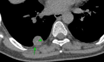
- Case 103: 53-years old - pleural mass in right medial cardiophrenic angle - solitary fibrous tumor of pleura
- Case 110: 84-years old - left chylothorax - pacemaker lead related left subclavian and innominate vein thrombosis causing thoracic duct obstruction
- Case 113: 74-years old - asbestos exposure - pleural plaques with rounded atelectasis
- Case of the Day 112: 82-years old - asbestos exposure - calcified pleural plaques
- Post 06: 44-years old - incidental nodule - adenocarcinoma with signs of visceral pleural invasion - jellyfish and multiple pleural tags
- Post 07: 77-years old with asbestosis pleural disease and tuberculosis
Chest Wall and Diaphragm
- Case 25: 45-years old - extrapleural masses - thoracic splenosis due to old diaphragmatic rupture
- Case 81: 57-years old - medial cardiophrenic angle mass - foramen of Morgagni hernia
- Case 82: 60-years old - type I sliding hiatus hernia
- Case 89: 72-years old man - diaphragmatic paralysis - vs eventration
- Case 93: 67-years old - Poland syndrome
- Case 99: 50-years old - intrathoracic rib
- Case of the Day 171: 56-years old - post-thoracoplasty and pneumonectomy sequelae 18 years later
Congenital and Developmental
- Case 13: 3-years old - congenital lobar emphysema
- Case 27: 18-years old - lung mass - sequestration
- Case 50: 43-years old - subcarinal bronchogenic cyst
- Case 61: 68-years old - venous aneurysms in lung
- Case 62: 41-years old - venous brachiocephalic aneurysm
- Case 63: 48-years old - unilateral absence pulmonary artery (UAPA)
- Case 65: 46-years old - Birt-Hogg-Dube (BHD) syndrome
- Case 72: 45-years old - Hermansky-Pudlak syndrome
- Case 93: 67-years old - Poland syndrome
- Case 99: 50-years old - intrathoracic rib
Infection
-
Tuberculosis
- Case 8: 14-years old - rupture of a large necrotic paratracheal mediastinal node into the lung - tuberculosis
- Case 17: 17-years old with hemoptysis - tuberculosis and both dense and soft centrilobular nodules
- Case 23: 60-years old - 40-pack years smoker - right upper lobe old granuloma
- Case 43: 21-years old - endobronchial and tracheal tuberculosis
- Case 69: 60-years old with mass-like calcific lesions
- Lecture: Tuberculosis vs sarcoidosis
- Case 84: 30-years old - classic miliary tuberculosis
- Lecture: Lung cancer screening in a TB endemic country
- Case 97: 38-year old - miliary tuberculosis presenting as ARDS
- Case 101 - 35-years old - miliary tuberculosis coexisting with infectious bronchiolitis and tree-in-bud lesions - disseminated tuberculosis
- Case 102: 76-years old - tracheobronchial tuberculosis - left main bronchus narrowing
- Lecture - Lung cancer screening in a TB endemic country - a radiologist's perspective
- Case 112 - 37-years old - hemoptysis and post-tuberculosis lung disease - PTLD - complication - fungal ball - chronic cavitary pulmonary aspergillosis - CCPA
- Post 07: 77-years old with asbestosis pleural disease and tuberculosis
- Case of the Day 187: 71 years old with miliary tuberculosis with a large broncholith in the right main bronchus
-
Fungal
- Case 16: 56-years old - Pneumocystis pneumonia (PCP) due to Pneumocystis jirovecii in a PLHA
- Case 20: 61-years old - chronic cavitary aspergillosis - scab sign
- Case 21: 61-years old - cryptococcus lung nodules
- Case 75: 51-years old - asthma - allergic bronchopulmonary aspergillosis (ABPA)
- Case 112: 37-years old - hemoptysis and PTLD - fungal ball in a cavity - CCPA
- Snippet 23: Allergic bronchopulmonary aspergillosis (ABPA) - Update of radiology subtypes
- Case of the Day 066: 60-years old - RA-ILD with chronic cavitary pulmonary aspergillosis (CCPA)
- Case of the Day 092: 44-years old - PCP/PJP in a PLHA
- Case of the Day 093: RA-Sjogren ILD with a variant UIP pattern with superimposed CCPA as a complication
- COD 137: 29-years old - chronic cavitary pulmonary aspergillosis - CCPA with "scab-like" sign
- Post 01: 59-years old with indeterminate fibrosing ILD but with likely immune-dysregulation related ILD with new diffuse ground glass due to PCP/PJP
-
Bacterial
-
Parasitic
-
Viral
- Case 2: 69-years old man with COVID-19 - resolution of so-called "fibrosis" at 9 months follow-up
- Case 14: 70-years old man with COVID-19 - perfusion defects due to COVID angiopathy at 3 1/2 weeks
- Case 18: 51-years man with COVID-19 - 11 months follow-up
- Case 19: 55-years old with COVID-19 vasculopathy / angiopathy - 10 1/2 months follow-up
- Case 32: 69-years old man with COVID-19 - 16 1/2 months follow-up
- Case 38: 40-years old man with COVID-19 - 15 months follow-up of COVID changes and vasculopathy
- Case 56: 66-years old - COVID-19 fibrosis - UIP-like
- Case 64: 70-years old - H1N1 lung involvement
- Case 100: 61-years old - Covid-19 related fibrosis - UIP like - plus review of Covid-19 lung changes 4 years later
- Case of the Day 118: 68-yrs old with post-COVID-19 residual lung abnormalities
- Case of the Day 196: 4 patients with different manifestations of COVID-19 from resolution to progressive fibrosis - 6 years on, the legacy of COVID-19
Neoplasms
-
Malignant
- Case 4: 70-years old with carcinoma breast - lymphangitis carcinomatosis
- Case 7: 75-years old man - UIP/IPF with a complication - lung nodule - squamous cell carcinoma
- Case 9: 64-years old - non-resolving consolidation - invasive mucinous adenocarcinoma
- Case 22: 64-years old - growing subsolid nodule - adenocarcinoma
- Case 40: 49-years old - primary tracheal chondrosarcoma
- Case 41: 78-years old - non-resolving opacity - invasive mucinous adenocarcinoma
- Case 42: 15-years old - prevascular space mass - lymphoma
- Case 86: 55-years old - growing cavitated lung nodule - adenocarcinoma metastasis
- Snippet 18: The jellyfish and multiple pleural tag signs to predict visceral pleural invasion in small lung cancers
- Case 115: 36-years old - lymphangitis from signet call adenocarcinoma
- Case of the Day 164: 66-years old with a Pancoast tumor - carcinoma
- Post 06: 44-years old - incidental nodule - adenocarcinoma with signs of visceral pleural invasion - jellyfish and multiple pleural tags
-
Benign
- Case 31: 54-years old - incidental lung nodule - pulmonary sclerosing pneumocytoma
- Case 52: 54-years old - solitary nodule with cyst - neuroendocrine tumor - carcinoid
- Case 53: 18-years old - lung mass with calcification - inflammatory myofibroblastic tumor
- Case 103: 53-years old - pleural mass in right medial cardiophrenic angle - solitary fibrous tumor of pleura
- Case 104: 69-years old - lung hamartoma
- Case 109: Two patients with typical nodules suggestive of well-differentiated neuroendocrine tumor on CT and DOTA-PET
- Post 05: 56-years old - incidental nodule - chondroid hamartoma
-
Lung Cancer Screening
- High risk CT imaging features in lung cancer screening - lecture
- Case 23: 60-years old - 40-pack years smoker - right upper lobe nodule - resolved using "mean" reconstructions to simulate a chest radiograph
- Lecture - Lung cancer screening in a TB endemic country
- Lecture - Lung cancer screening in a TB endemic country - a radiologist's perspective
Connective Tissue Diseases and Vasculitides and Sarcoidosis
-
General
-
Sarcoidosis
- Case 29: 58-years old - sarcoidosis over 18 years
- Case 68: 47-years old - Stage II sarcoidosis - nodules and nodes
- Snippet 15: CT scan findings in sarcoidosis
- Lecture: Tuberculosis vs sarcoidosis
- Lecture: Imaging of Sarcoidosis
- Case of the Day 149: 40-years old with subtle lung findings and adenopathy - sarcoidosis
- Post 03: 41-years old with Stage III sarcoidosis - 1 year follow-up showing improvement with "melted sugar/cotton candy/tinted" sign of partial regression
- Case of the Day 172: 49-years old - perivascular, fissural and subpleural nodules with adenopathy - classic stage II sarcoidosis
-
Rheumatoid Arthritis
- Case of the Day 066: 60-years old - rheumatoid arthritis (RA) ILD with a usual interstitial pneumonia (UIP) pattern with variant fibrosis with superimposed CCPA
- Case of the Day 093: Why AI is the only solution to our chest X-ray hyposkillia - Rheumatoid arthritis-Sjogren ILD with a variant UIP pattern with superimposed CCPA as a complication
- Case of the Day 099: 61-years old - multiple waxing waning lung nodules - rheumatoid arthritis - rheumatoid nodules
- Post 04: 64-years old smoker with RA - RA, RA-ILD, AEF and CPFE
-
Scleroderma
- Case 39: 27-years old - scleroderma ILD - secondary pleuroparenchymal fibroelastosis (PPFE)
- Case 58: 27-years old - scleroderma ILD - progressive pulmonary fibrosis
- Case 94: 24-years old - scleroderma ILD - early/subtle disease
- Case of the Day 116: 31-yrs old with scleroderma and a mixed BIP/NSIP/CiOP pattern - mixed patterns in connective tissue disease ILDs
- Case of the 117: 51-yrs old in 2012 with a mixed progressive fibrotic NSIP/OP/DPO patter, diagnosed to have scleroderma/MCTD 13 years later
- Case of the Day 174: 33-years old with scleroderma showing an NSIP pattern along with FANO - faint amorphous nodular opacification
-
Sjogren's
-
Vasculitis
- Case 44: 33-years old - ANCA-associated vasculitis and diffuse alveolar hemorrhage
- Case 83: 51-years old - progressive masses and nodules - GPA (granulomatosis with polyangitis)
- Snippet 30: Vasculitis
- Post 08: 52-years old - multifocal peribronchovascular conslidation - ANCA associated vasculitis
- Case of the Day 194: 37-years old with variable vessel vasculitis - Behcet's disease
Exposure & Injury including Iatrogenic and Post-Operative
(Smoking, Occupational, Biomass Gas) / Poisonings / Radiation & Medication Induced Injury)
-
Smoking
- Case 23: 60-years old - 40-pack years smoker - right upper lobe nodule - resolved using "mean" reconstructions to simulate a chest radiograph
- Snippet 12: Visual classification of centrilobular and paraseptal emphysema
- Case 47: 64-years old smoker - respiratory bronchiolitis (RB) and discussion of smoking related changes
- Snippet 13: Spectrum of smoking related changes in a single patient
- Case 48: 78-years old - combined pulmonary fibrosis and emphysema (CPFE)
- Case 67: 69-years old - combined pulmonary fibrosis and emphysema (CPFE) and the official statement from ATS/ERS/JRS/ALAT
- Lecture: REF-Cafe Roentgen - smoking related lung diseases
- Snippet 27: Airspace Enlargement with Fibrosis (AEF)
- Post 04: 64-years old smoker with RA - RA, RA-ILD, AEF and CPFE
-
Air Pollution / Biomass Gas
-
Occupational/Pneumoconiosis
- Case 30: 51-years old - classic silicosis
- Case 45: 51-years old - eggshell calcification due to silicosis
- Case 88: 48-years old - flour mill worker silicosis
- Case 113: 74-years old - asbestos exposure - pleural plaques with rounded atelectasis
- Case of the Day 095: 55-years old - asbestos exposure - early/mild asbestosis with subpleural bands/lines
- Case of the Day 112: 82-years old - asbestos exposure - calcified pleural plaques
- Post 07: 77-years old with asbestosis pleural disease and tuberculosis
- Post 09: 60-years old flour-mill worker with variable-sized small nodules - silicosis, not miliary tuberculosis
-
Poisoning
-
Medication Induced Lung Injury (MIPI)
-
Radiotherapy-Associated Lung Injury
-
Post-operative
REF-Cafe Roentgen Thoracic Thursday Lecture Series - 2023
- Lecture 01 - 2023 06 15 - Fibrosing ILDs
- Lecture 02 - 2023 06 22 - Fibrosing ILDs - contd, Technique & Introduction to Non-Fibrosing ILDs
- Lecture 03 - 2023 06 29 - Non-fibrosing ILDs
- Lecture 04 - 2023 07 13 - Non-ILD Diffuse Lung Diseases - Bronchiectasis, Emphysema, Constrictive Bronchiolitis
- Lecture 05 - 2023 07 20 - Centrilobular Nodule, Proliferative Bronchiolitis, etc
- Lecture 06 - 2023 08 03 - Smoking Related Lung Diseases
- Lecture 07 - 2023 08 10 - Diffuse Cystic Lung Disease
- Lecture 08 - 2023 09 07 - Solitary Pulmonary Nodule - I - >8mm
- Lecture 09 - 2023 09 14 - Solitary Pulmonary Nodule - II - PFN, Juxtapleural Nodule, Nodules in Smokers, LungRADS
- Lecture 10 - 2023 09 21 - Solitary Pulmonary Nodule - III - Subsolid Nodule
- Lecture 11 - 2023 10 19 - Non-Resolving Opacity
- Lecture 12 - 2023 10 26 - Mediastinum