COD 201 - A Nodule in a Smoker - What Now?
74-year old with a 25-pack years history of smoking
Case:
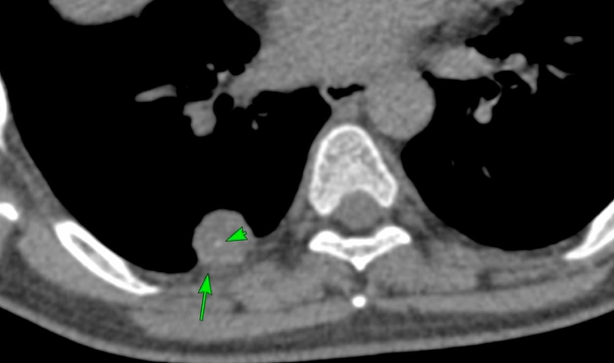
74-years old with a 25-pack years history of smoking. He had a lesion in the left ureter and came for imaging.
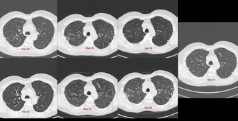
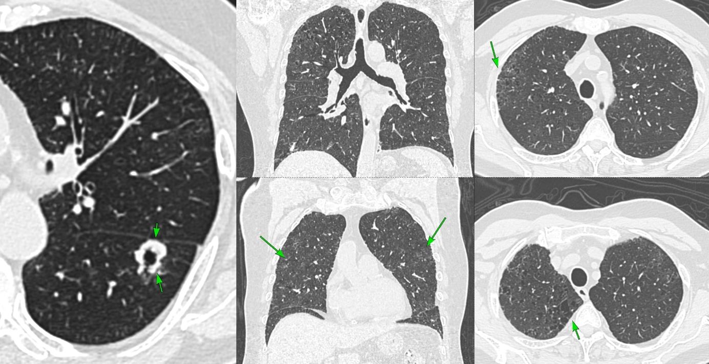
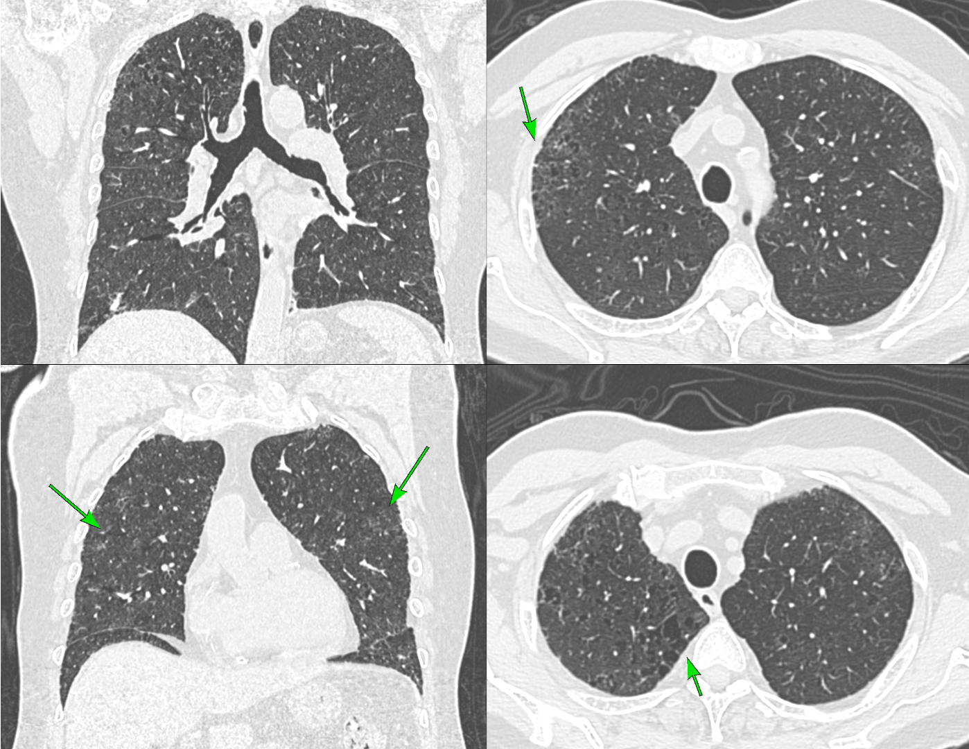
Smoking related changes (Fig. 1)
- Confluent centrilobular emphysema
- Subtle mild paraseptal emphysema
- Respiratory bronchiolitis
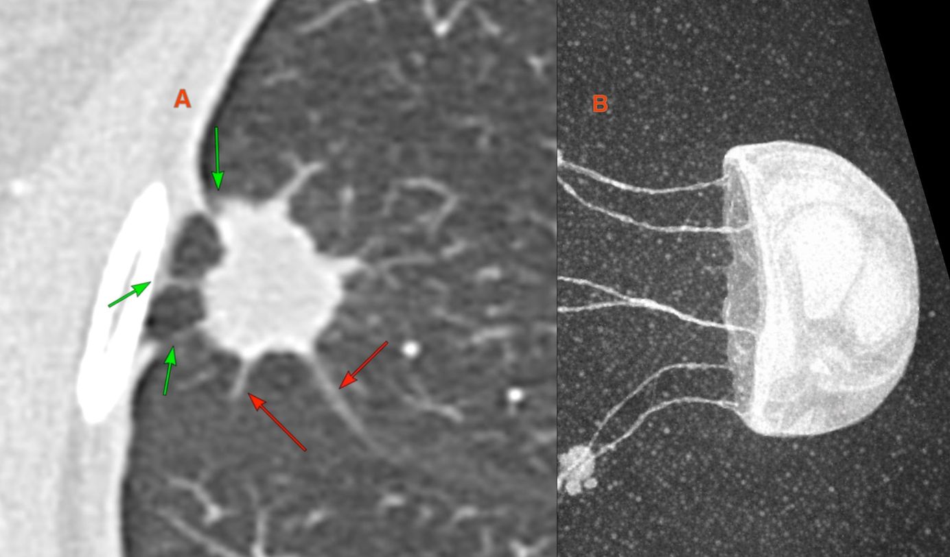
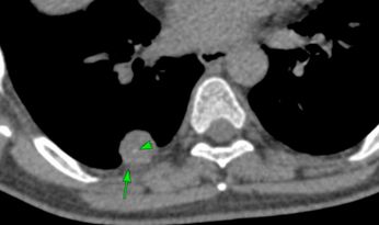
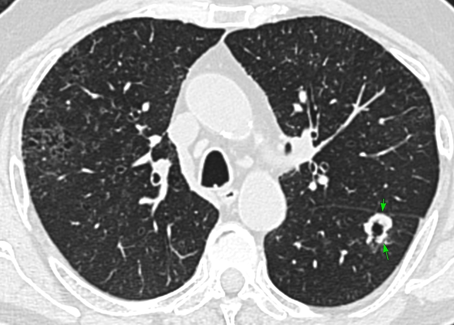
He also had a lung nodule
The video walks through how to read a smoker's CT in "smoker's protocol" mode.
- The spectrum of smoking-related lung changes
- Hunting for nodules with 5–7 mm MIPs
- Characterising what we find — a lobulated, cavitated nodule with subtle spiculation
- Applying Lung-RADS 2022 — categories 1, 2, 3, 4A, 4B, 4X
This post is for subscribers only
SubscribeAlready have an account? Log in