Post 11 - The Natural Evolution of Radiotherapy-Associated Lung Injury
39-years old with carcinoma breast
Case:
39-years old with carcinoma breast underwent surgery in Sep 24, followed by radiotherapy (RT) in Nov 24.
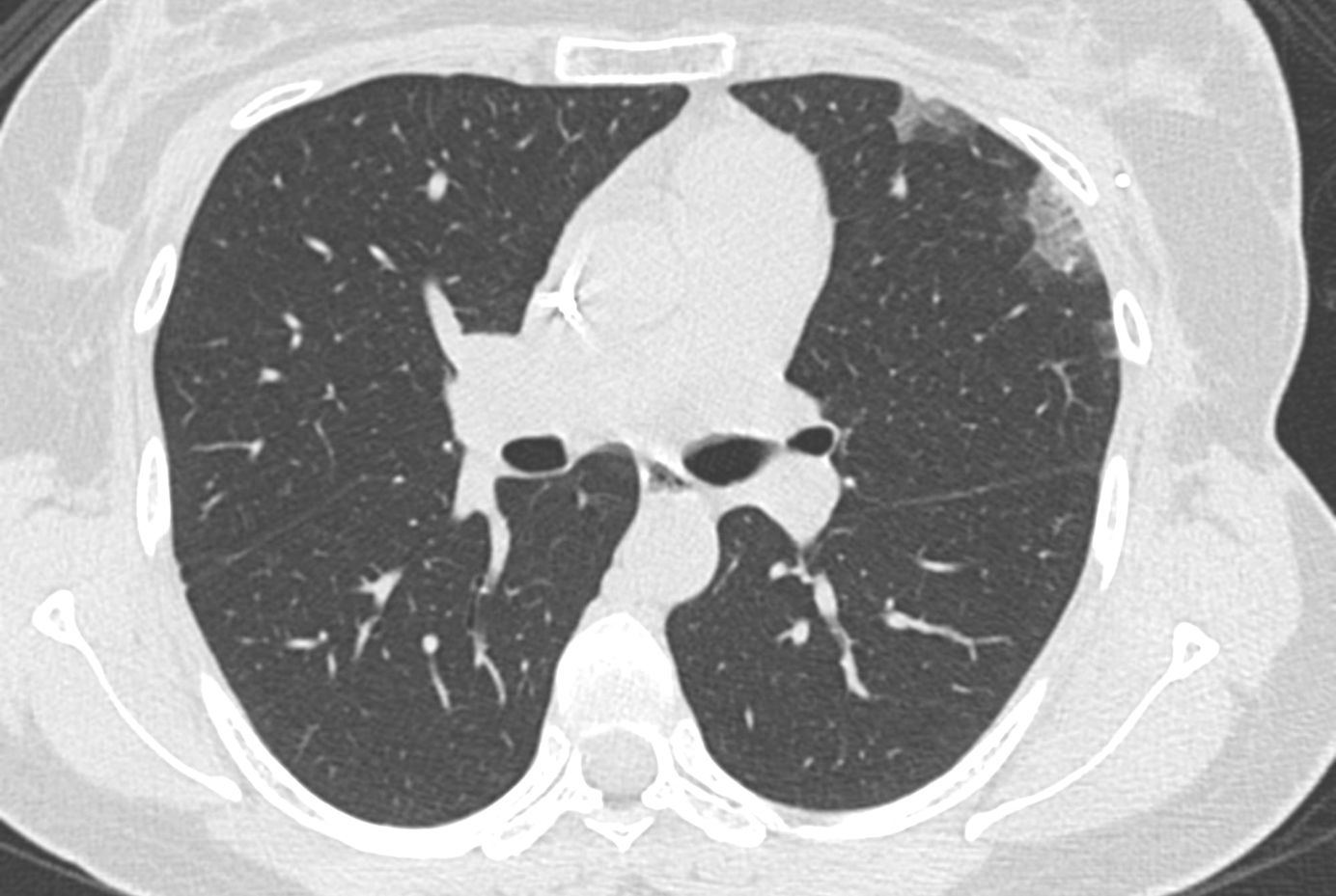
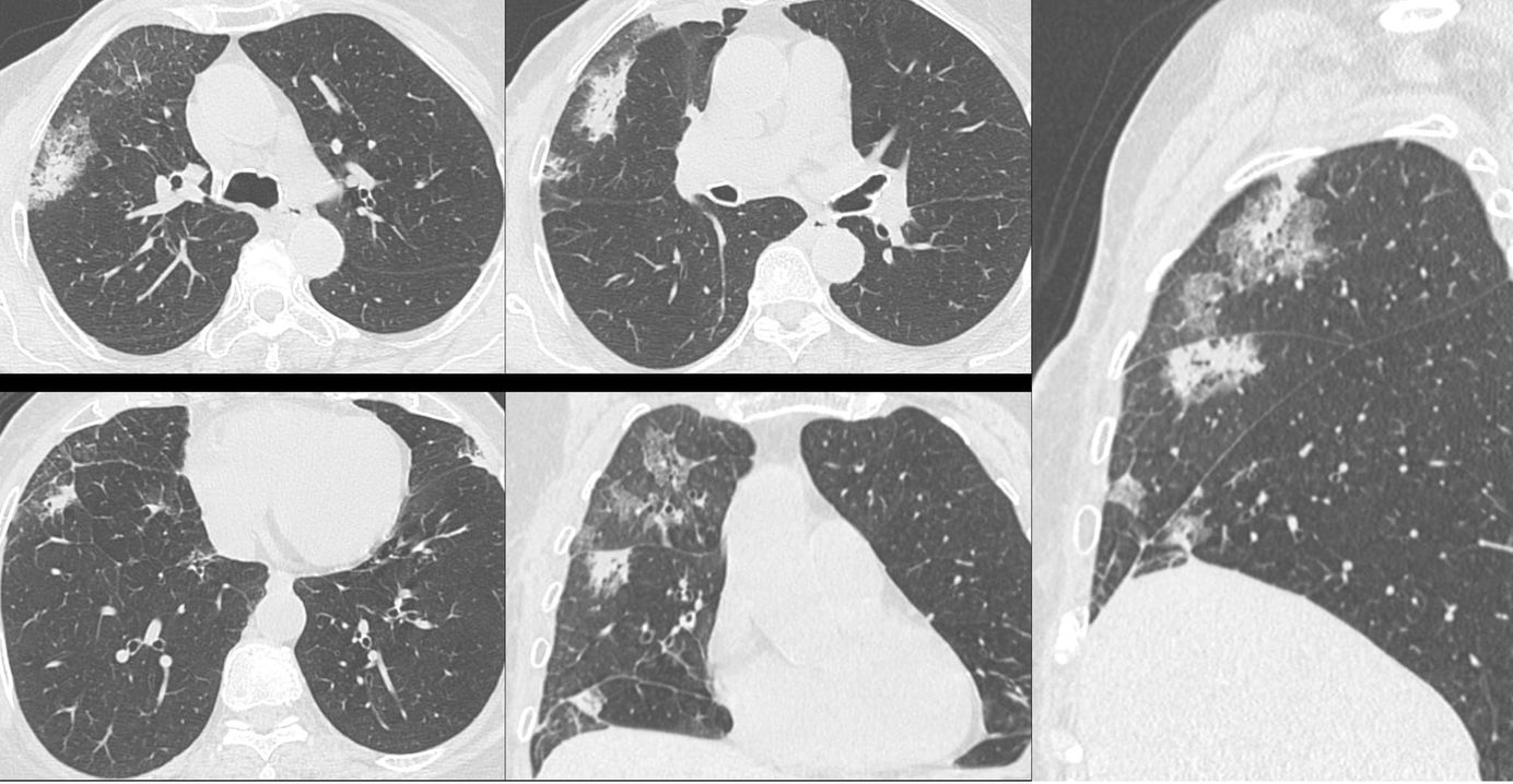
The CT chest (Fig. 1 - bottom row) obtained as part of a PET/CT in Jun 25, 7 months later shows areas of organizing pneumonia in the subjacent lung of the left upper lobe and lingula.

One year and 6 months later (Fig.1 - top row), the lesions are now fibrotic, presenting as curvilinear bands.
This is the natural progression of any acute lung injury from exudation to organization to healing (fibrosis vs complete regression) and for radiotherapy-associated lung injury, the timeline has been eloquently shown in this diagram from a recent Radiographics article (Fig. 2).

To know more about radiotherapy-associated lung injury, a recent snippet.

Previous Similar Posts


Index and Table of Contents
116 Cases with Videos
11 Cases with Posts
31 Snippets
16 Lectures
36 Cases of the Day (CODs)
New Series - ILD Patterns (3 Done)

New One-Time, Lifetime Subscription
Please check out this page to see the changes in subscription models.

Previous Posts:


CT Chest Review Newsletter
Join the newsletter to receive the latest updates in your inbox.
