Post 08 - Multifocal Peribronchovasular Consolidation
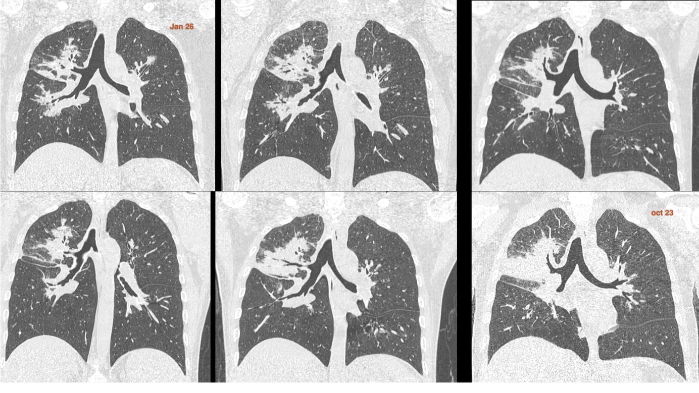
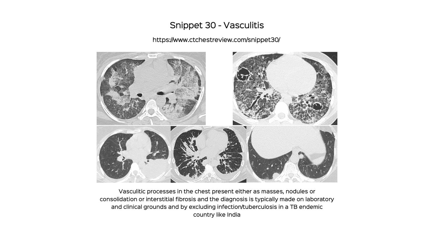
The pattern of peribronchovascular consolidation and its differentials
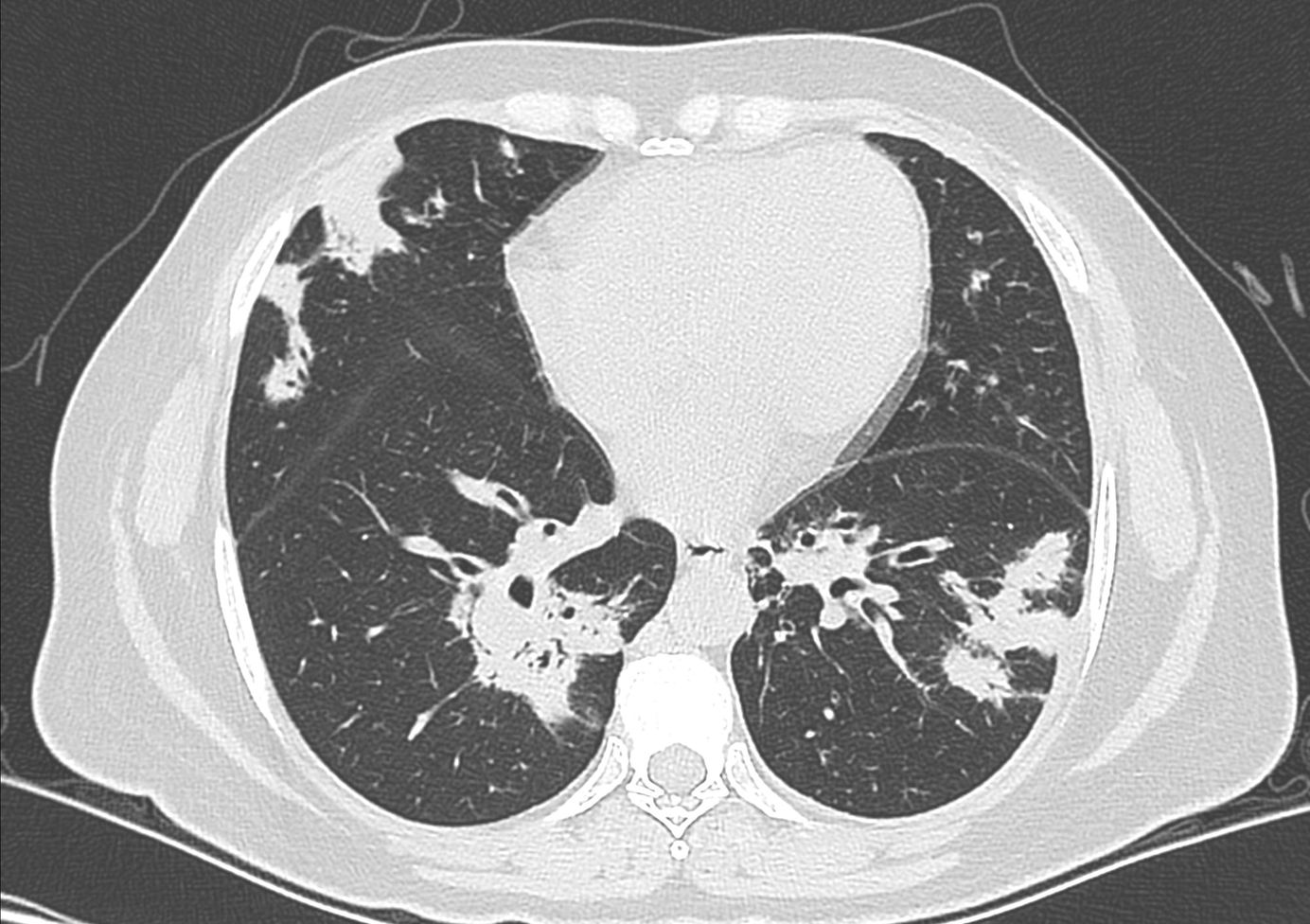
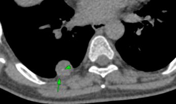
52-years old came for a CT chest.
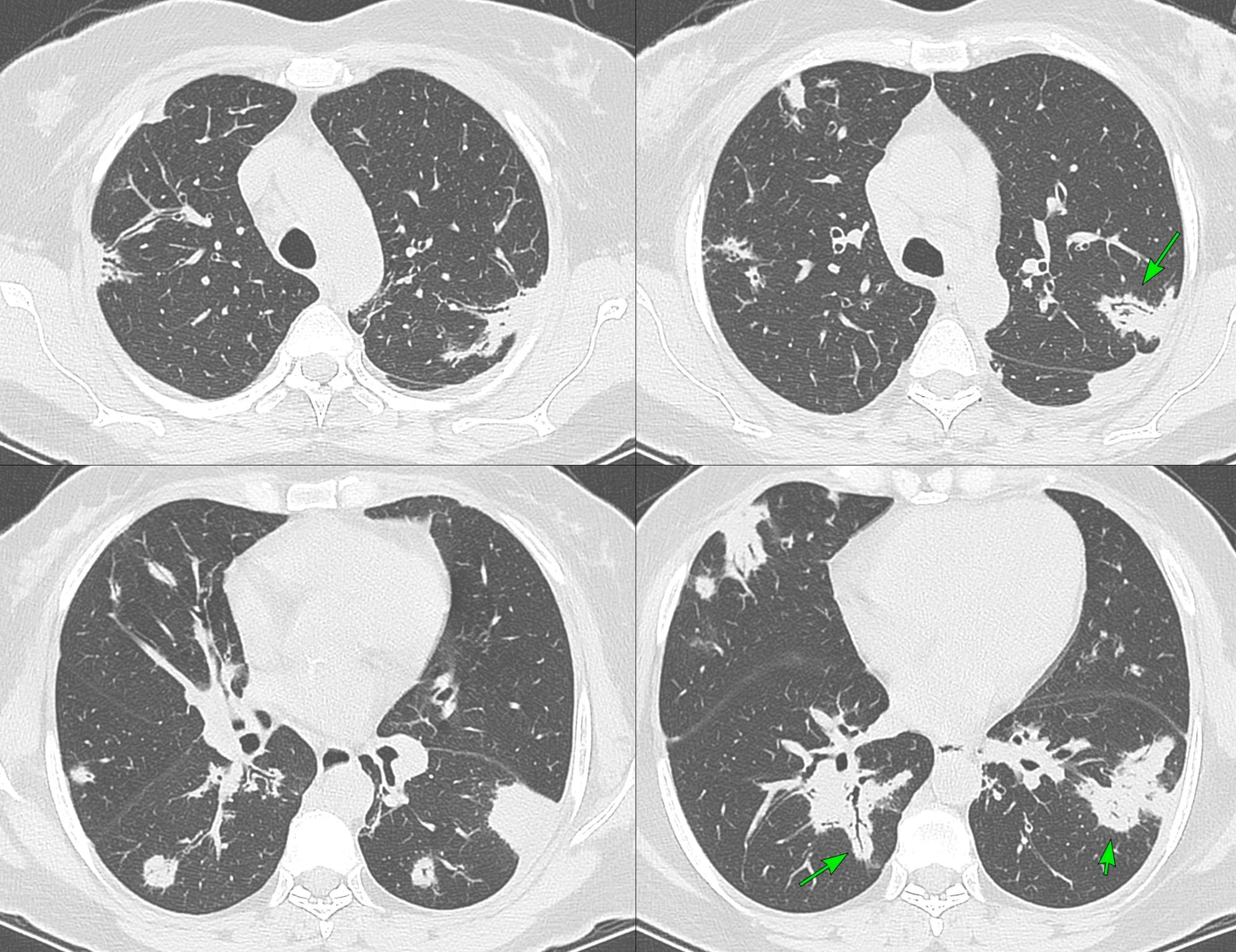
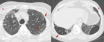
The CT shows peribronchovascular areas of consolidation with air bronchograms (arrows).

This is characteristic asbestos exposure related pleural disease.
This is an organizing pneumonia (OP) pattern, which needs to be further investigated for etiology.
However, this type of OP pattern itself has multiple differentials including
- Lymphoma
- IgG4-related disease
- Vasculitis
- Sarcoidosis
This patient was ANCA-positive and had a sural nerve biopsy showing lymphocytic vasculitis.
This pattern is hence consistent with ANCA-associated vasculitis presenting with peribronchovascular areas of consolidation.
More about Vasculitis


Index and Table of Contents
116 Cases with Videos
8 Cases with Posts
30 Snippets
16 Lectures
31 Cases of the Day (CODs)
New Series - ILD Patterns (2 Done)

New One-Time, Lifetime Subscription
Please check out this page to see the changes in subscription models.

Previous Posts:


CT Chest Review Newsletter
Join the newsletter to receive the latest updates in your inbox.
