Case 69: What Ails Chest Imaging Training?
It helps to follow the first principles of interpretation
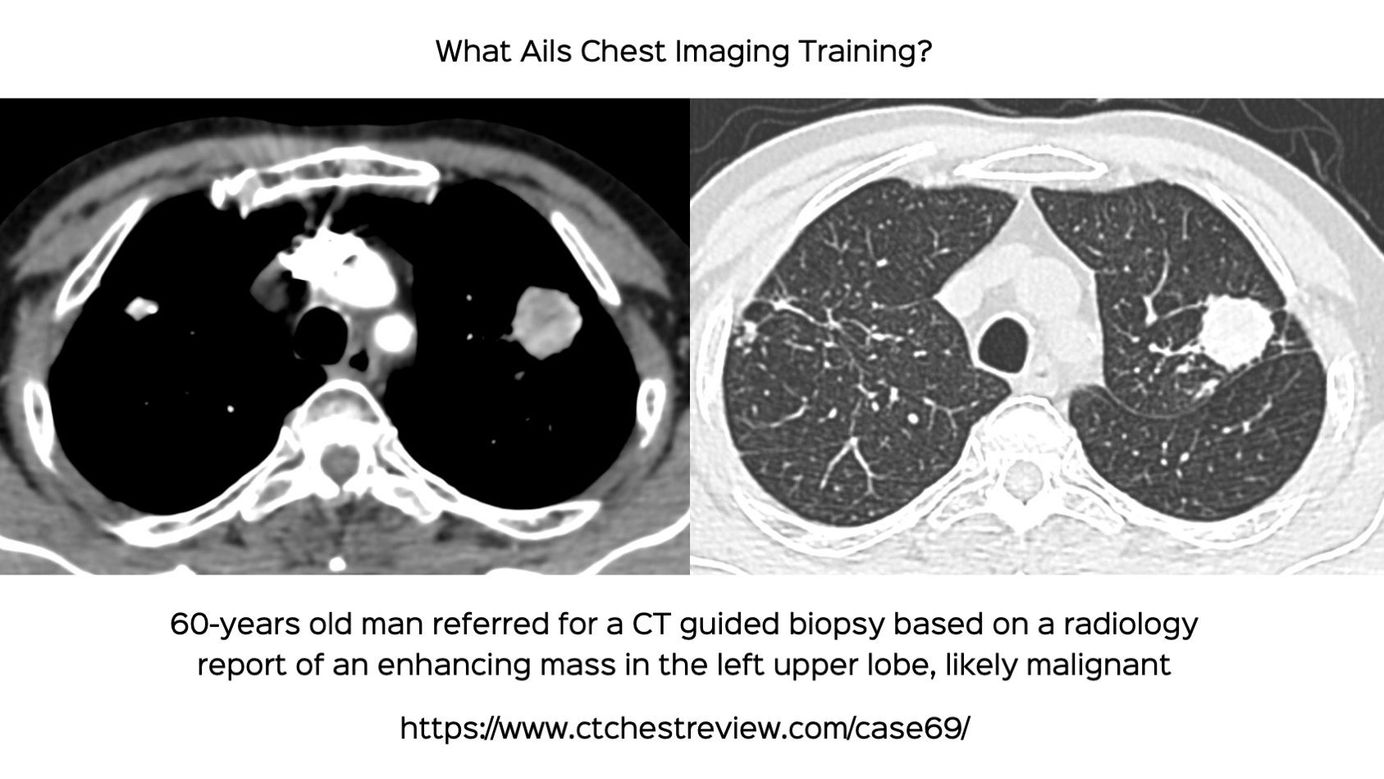
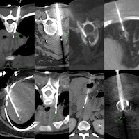
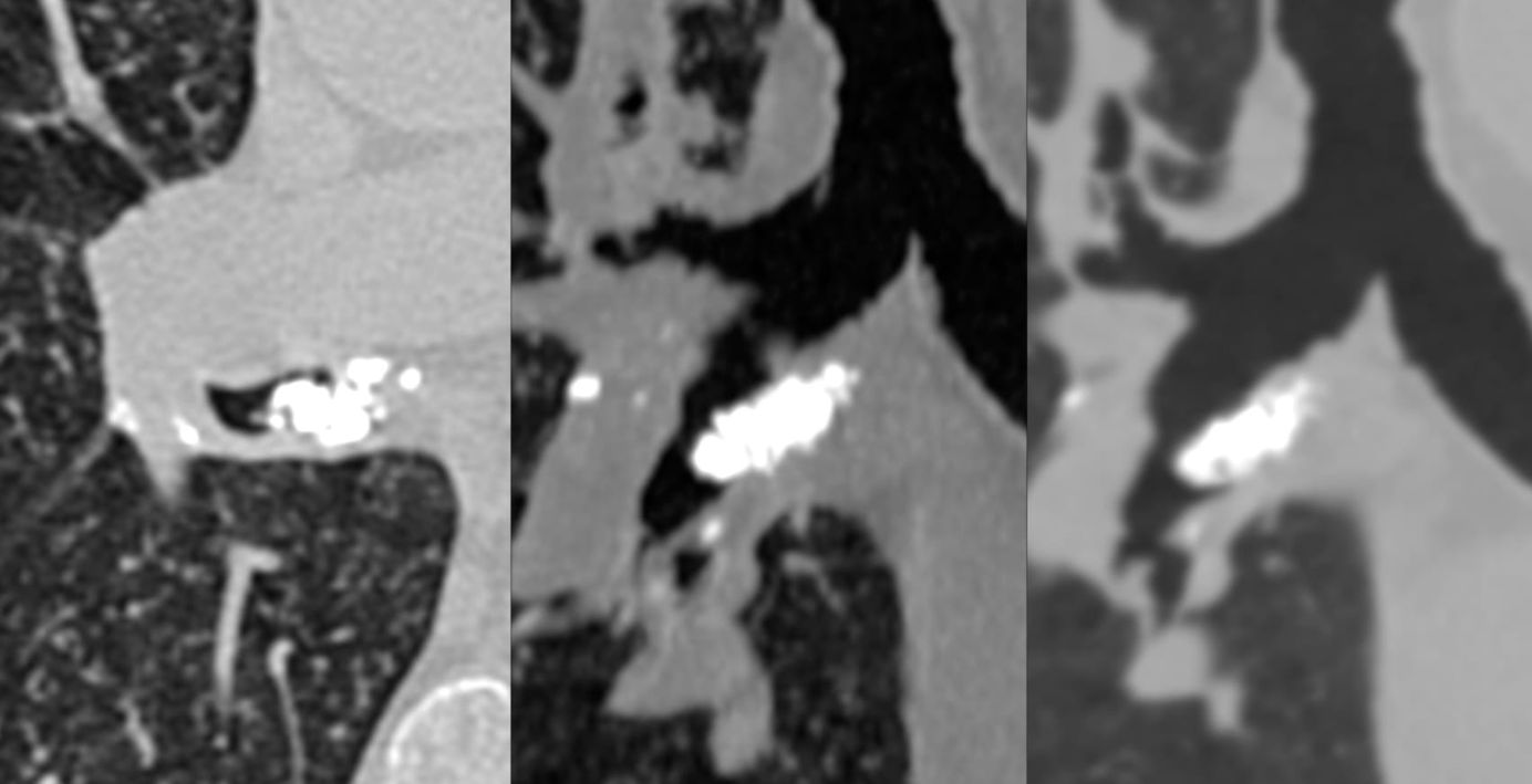
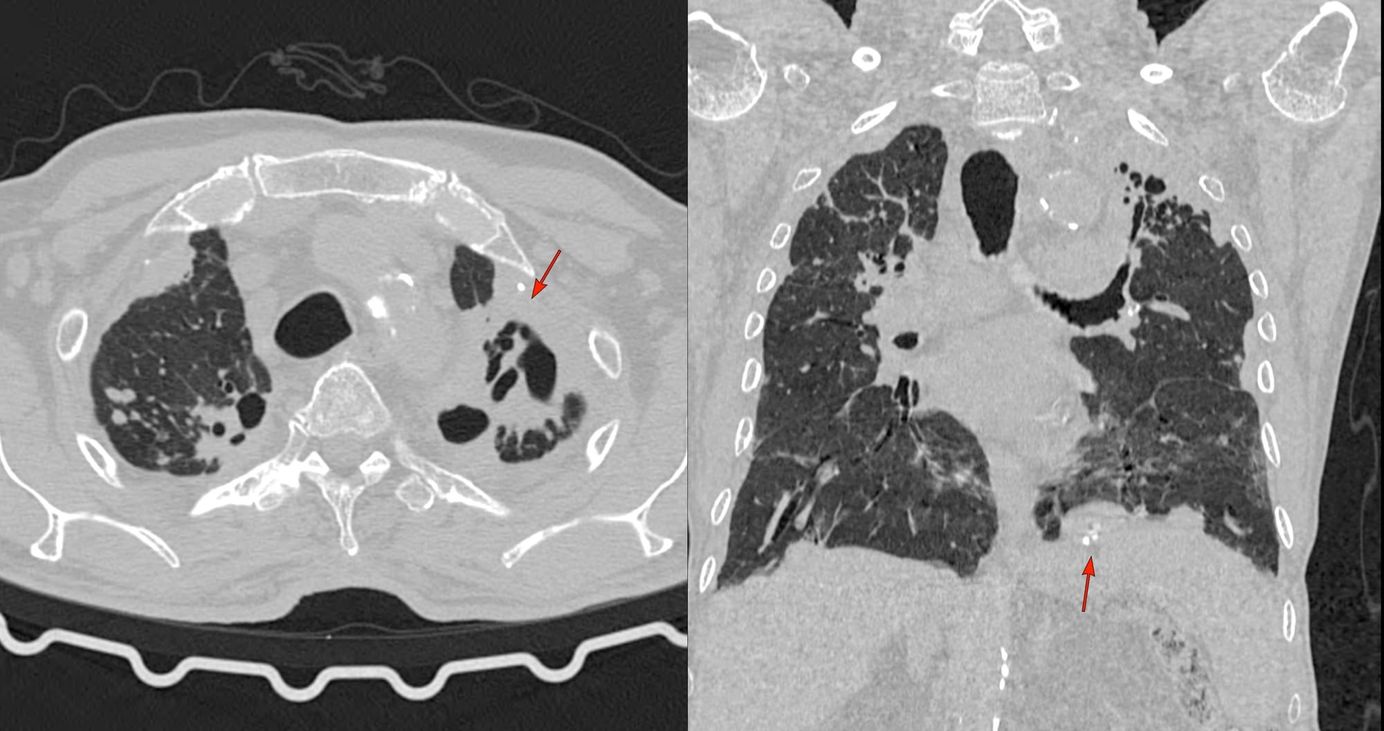
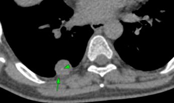
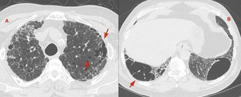
This is a 60-years old man who was referred for a CT guided biopsy of a left upper lobe lung mass, that turned out not to be so. The reporting person had not bothered to look at the plain scan and to check for calcification / enhancement, etc.
This is one of the first principles of CT scan interpretation. Look at the plain scan to check for calcification and enhancement.
The video runs through the case, other similar cases, discusses the first principles of interpretation and the results of the recent AI paper on diagnosing active TB with respect to the performance of India-based radiologists compared to AI and US-based radiologists.
Table of Contents
Other Cases
 Bhavin Jankharia
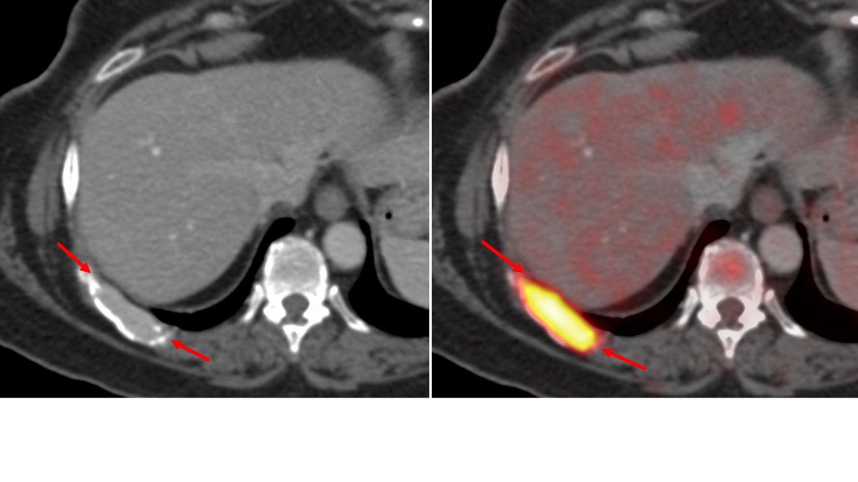

CT Chest Review Newsletter
Join the newsletter to receive the latest updates in your inbox.