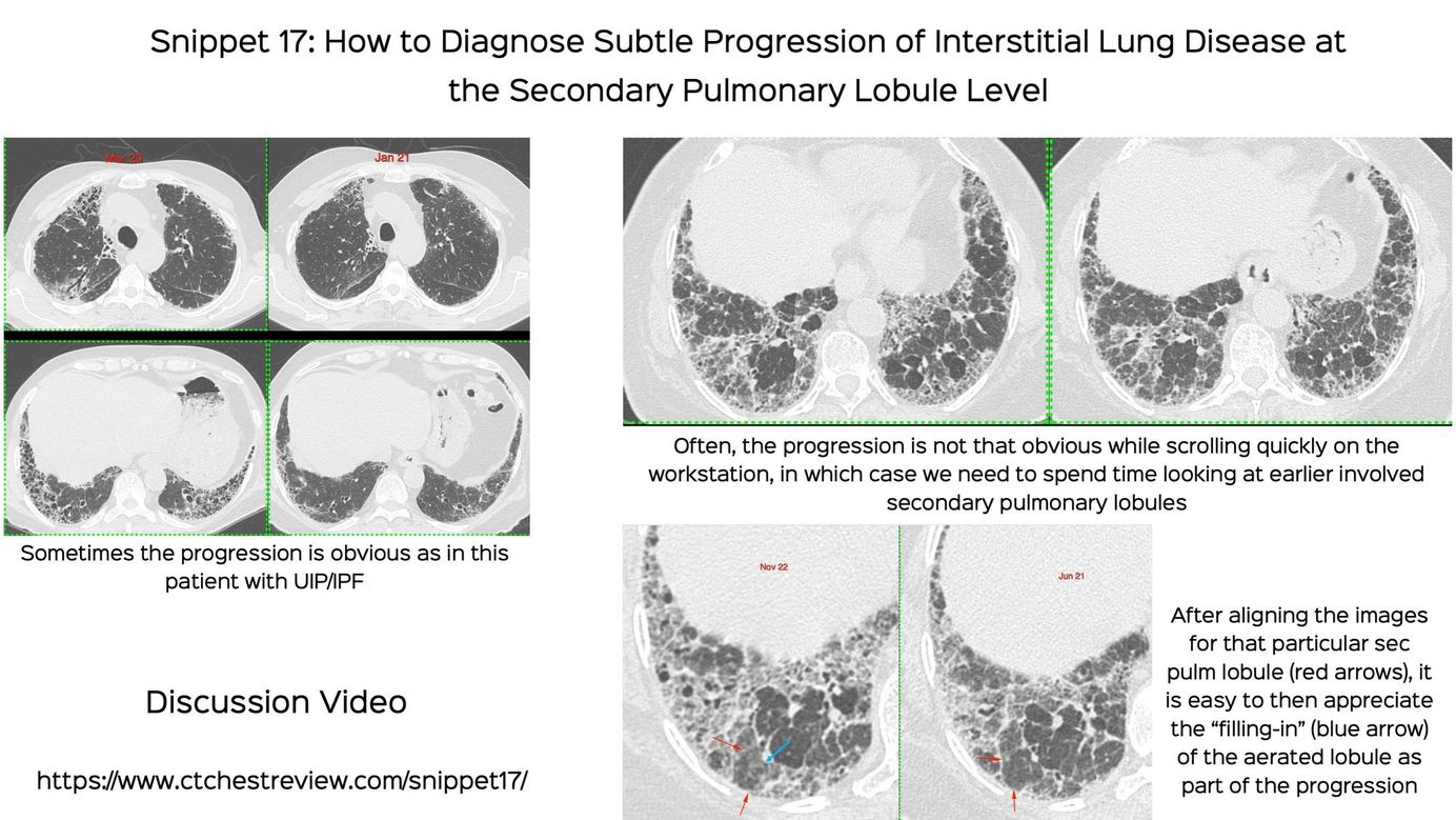
Snippet 17: How to Diagnose Subtle Progression of Interstitial Lung Disease at the Secondary Pulmonary Lobule Level
How to look for progression at the secondary pulmonary lobule level, if it is not obvious on routine scrolling
Table of Contents
Index and Table of Contents
Technical and Practice Issues * Case 11: 62-years old misdiagnosed to have interstitial lung disease - mid-inspiratory and expiratory scans * Snippet 03: Radiation risk and CT chest * Case 23: 60-years old - 40-pack years smoker - right upper lobe nodule - resolved using “mean” reconstructions…
Text
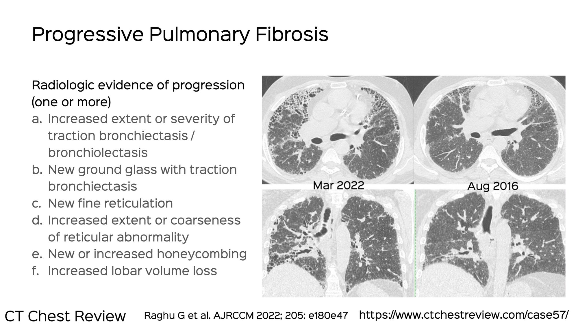
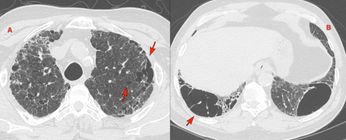
As seen in this figure depicting the criteria for progressive pulmonary fibrosis in a patient of fibrotic hypersensitivity pneumonitis from 2016 to 2022, usually it is quite easy to diagnose progression when scrolling through the images on the workstation.
Sometimes however, the scans look almost the same. In that case, it is important to spend a little more time looking at the uninvolved areas to see if there is "filling-in" of the previously uninvolved secondary pulmonary lobules, which would then suggest progression.
The video describes this process in detail.
This post is for paying subscribers only
SubscribeAlready have an account? Log in