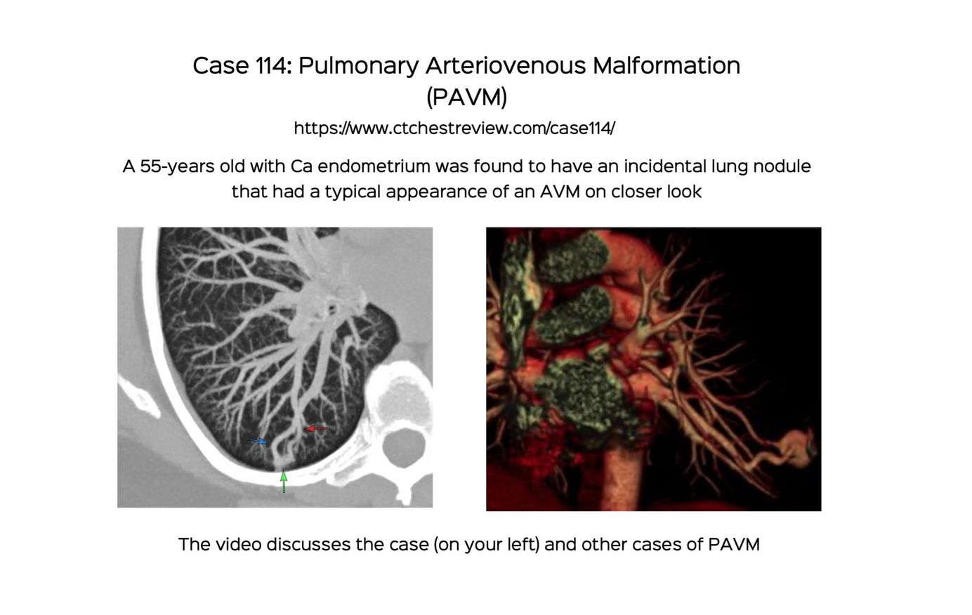
Case 114: Pulmonary Arteriovenous Malformation (PAVM)
Pulmonary AVMs are typically easy to diagnose on plain CT. Contrast CT helps with confirmation and planning. Embolization is the typical mode of treatment, even if the patient is asymptomatic.
New One-Time, Lifetime Subscription
Please check out this page to see the changes in subscription models.
Payment
To make the site more accessible, we have decided to remove the yearly subscription and keep only a one-time, lifetime payment to get access to all content at www.ctchestreview.com and www.ctbiopsy.com. These sites are linked and hence one payment gives access to both sites, but the
Current Post
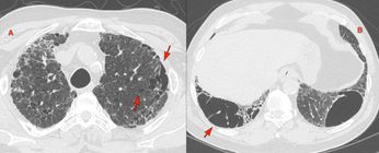
A 55-years old with carcinoma endometrium was referred for an opinion for a lung nodule diagnosed on the PET/CT.
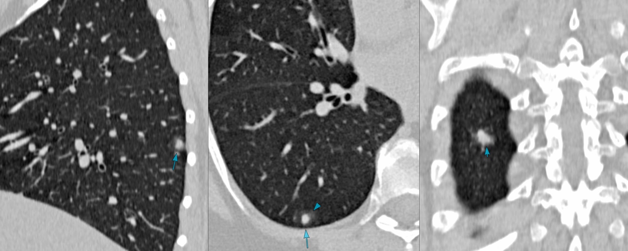
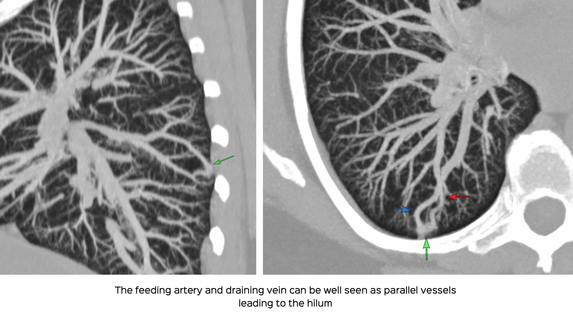
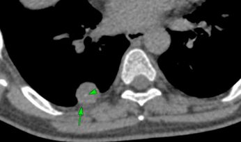
A closer look showed linear vessels leading up to the nodule and MIP images showed a classic AVM.
The video discusses the case and the current knowledge base of PAVMs along with other similar cases
This post is for paying subscribers only
SubscribeAlready have an account? Log in

)