Lecture: CT of Hypersensitivity Pneumonitis
CT scan findings in hypersensitivity pneumonitis, fibrotic and nonfibrotic
Table of Contents
Index and Table of Contents
Technical and Practice Issues * Case 11: 62-years old misdiagnosed to have interstitial lung disease - mid-inspiratory and expiratory scans * Snippet 03: Radiation risk and CT chest * Case 23: 60-years old - 40-pack years smoker - right upper lobe nodule - resolved using “mean” reconstructions…
Previous Case
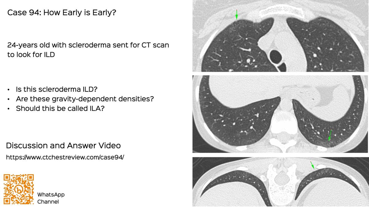
Case 94: How Early is Early...
What do these findings in a patient with scleroderma signify?
Current Lecture
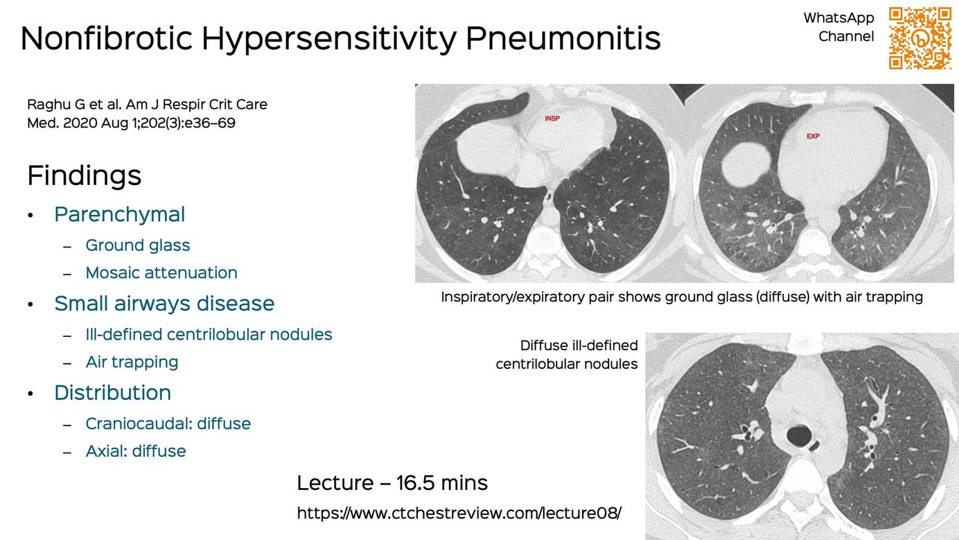
This is a lecture I gave earlier this week. It is a 16/12 minutes short lecture on CT scan in hypersensitivity pneumonitis.
I discuss the signs of nonfibrotic hypersensitivity pneumonitis followed by a case showing progression from nonfibrotic to fibrotic hypersensitivity pneumonitis and then discuss the signs of fibrotic hypersensitivity pneumonitis along with progressive pulmonary fibrosis (PPF).
This post is for subscribers only
SubscribeAlready have an account? Log in