COD 193 - Not Every Lung Infarct is Thromboembolic
74-years old with an infarct
Case:
74-years old man presented with abdominal pain and he was diagnosed with adenocarcinoma pancreas with liver metastases from a CT guided liver lesion biopsy after a PET/CT.
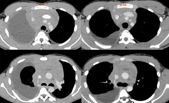
The chest showed
- Ill-defined lesion in the right infrahilar region
- Encasing right inferior pulmonary vein
- Rt pleural effusion
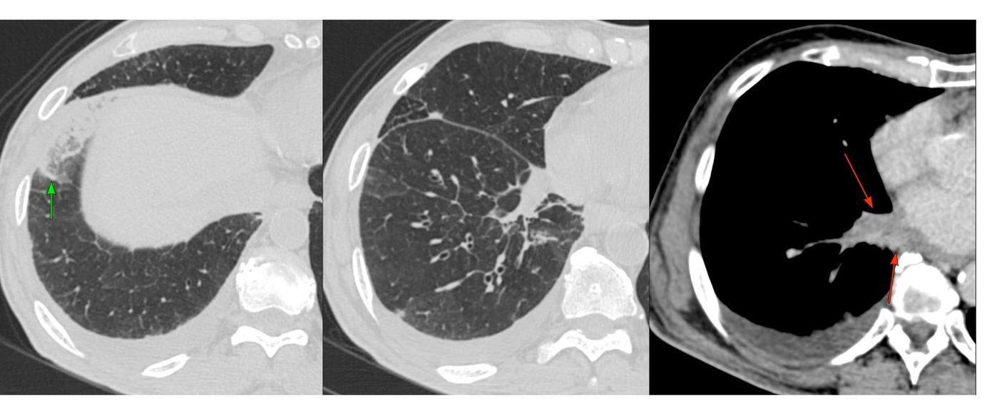
- Bubble lucencies in a focal lung lesion in the anterior and lateral basal segments of the right lower lobes
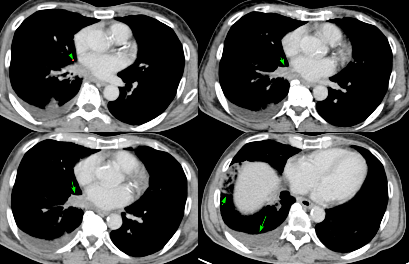
These were infarcts.
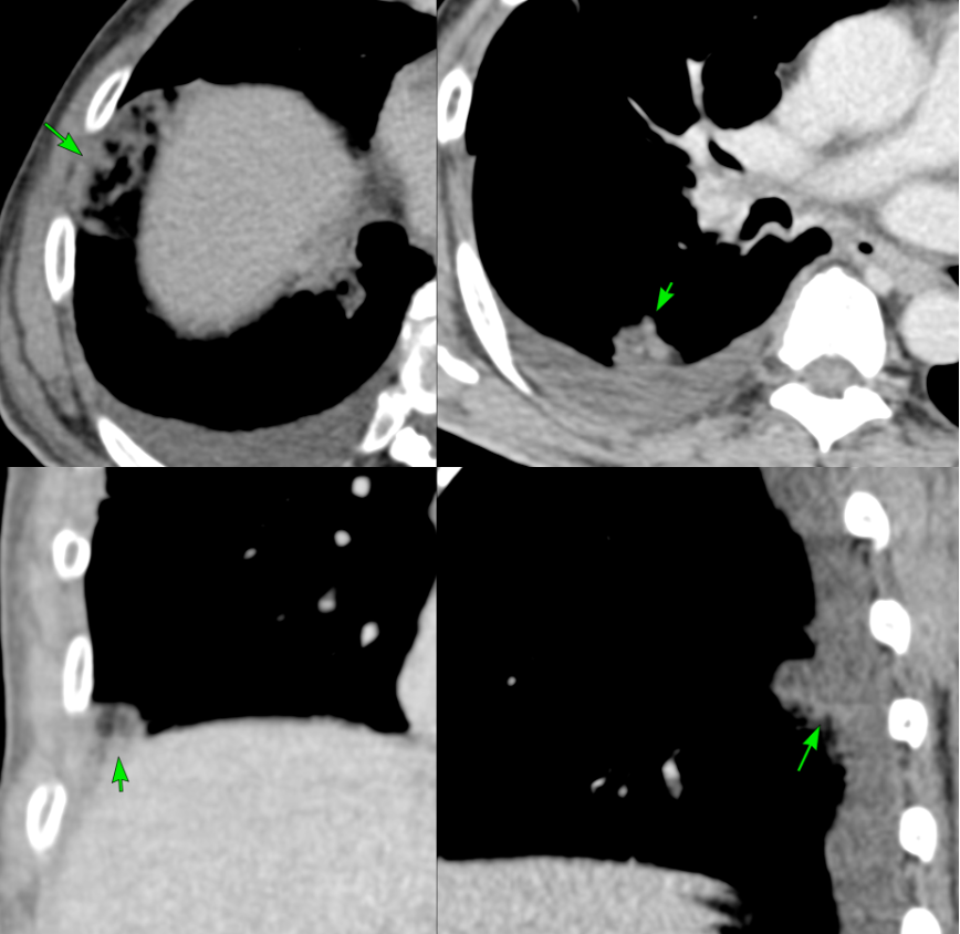
His pulmonary arteries were normal, but the pulmonary vein was involved.
This post is for paying subscribers only
SubscribeAlready have an account? Log in