COD 189 - Idiopathic Pleuroparenchymal Fibroelastosis (iPPFE) - Volumes, Flat Chest and Meandering Trachea
All the findings that go into making a diagnosis of iPPFE
Index and Table of Contents
116 Cases with Videos
5 Cases with Posts
30 Snippets
16 Lectures
31 Cases of the Day (CODs)
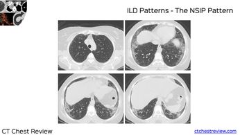
New Series - ILD Patterns (2 Done)
Index and Table of Contents
Technical and Practice Issues * Case 11: 62-years old misdiagnosed to have interstitial lung disease - mid-inspiratory and expiratory scans * Snippet 03: Radiation risk and CT chest * Case 23: 60-years old - 40-pack years smoker - right upper lobe nodule - resolved using “mean” reconstructions…
New One-Time, Lifetime Subscription
Please check out this page to see the changes in subscription models.
Payment
To make the site more accessible, we have decided to remove the yearly subscription and keep only a one-time, lifetime payment to get access to all content at www.ctchestreview.com and www.ctbiopsy.com. These sites are linked and hence one payment gives access to both sites, but the
Case
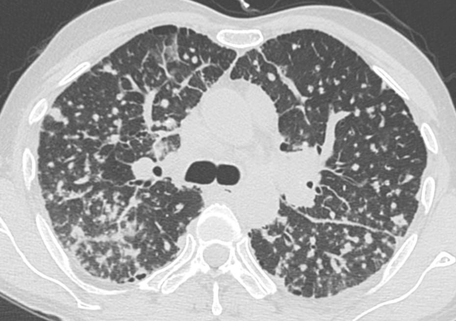
37-years old with gradually progressive dyspnea.
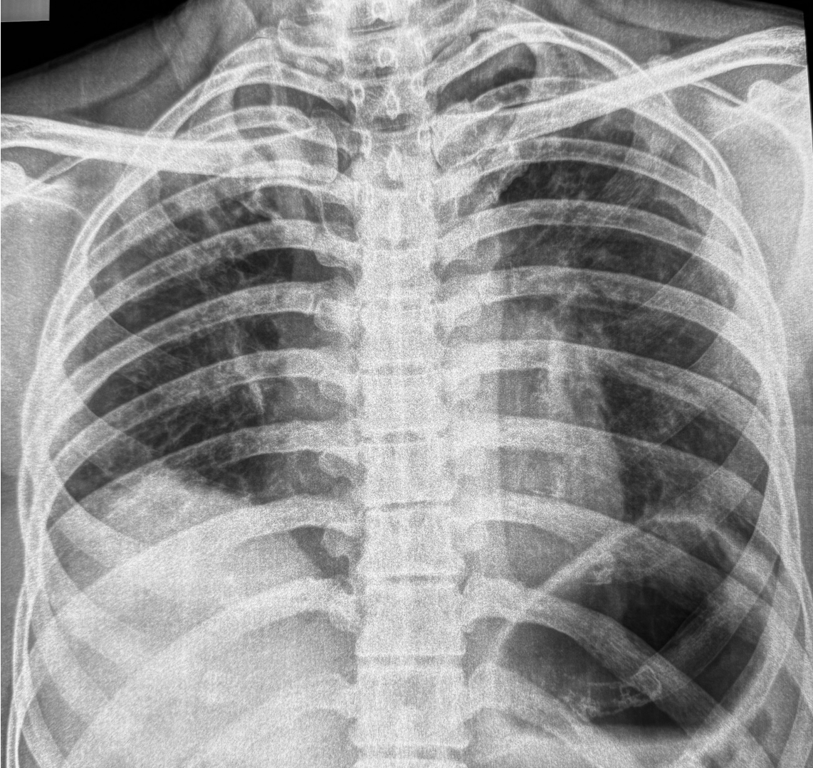
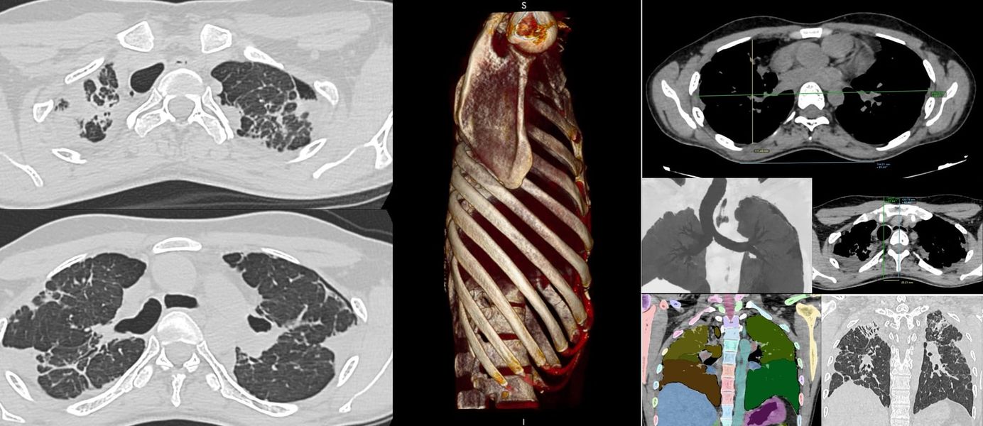
Upper lobe reticular opacities with volume loss, extending below tracheal bifurcation, small left pneumothorax.
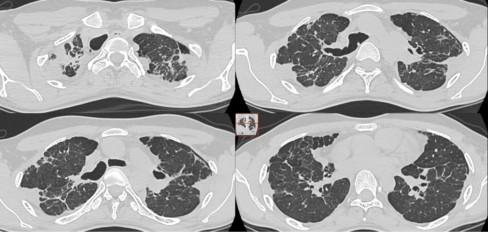
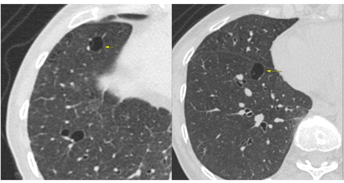
CT images show classic findings of pleuroparenchymal fibroelastosis (PPFE), with no other associated etiology, hence idiopathic - iPFFE.
The video describes all the additional findings that often go hand in hand as in
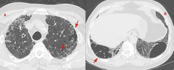
- Playthorax - flat chest - progression of platythorax associated with poorer prognosis
2l Anterior and downward rotation of the ribs - associated with poorer prognosis - Reduced upper lobe lung volumes
- Deep suprasternal notch (not present in our patient)
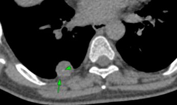
- Tracheal deviation and meandering trachea.
To know each time a new post is up
This post is for paying subscribers only
SubscribeAlready have an account? Log in
%20%20-%20Volumes%2C%20Flat%20Chest%20and%20Meandering%20Trachea)