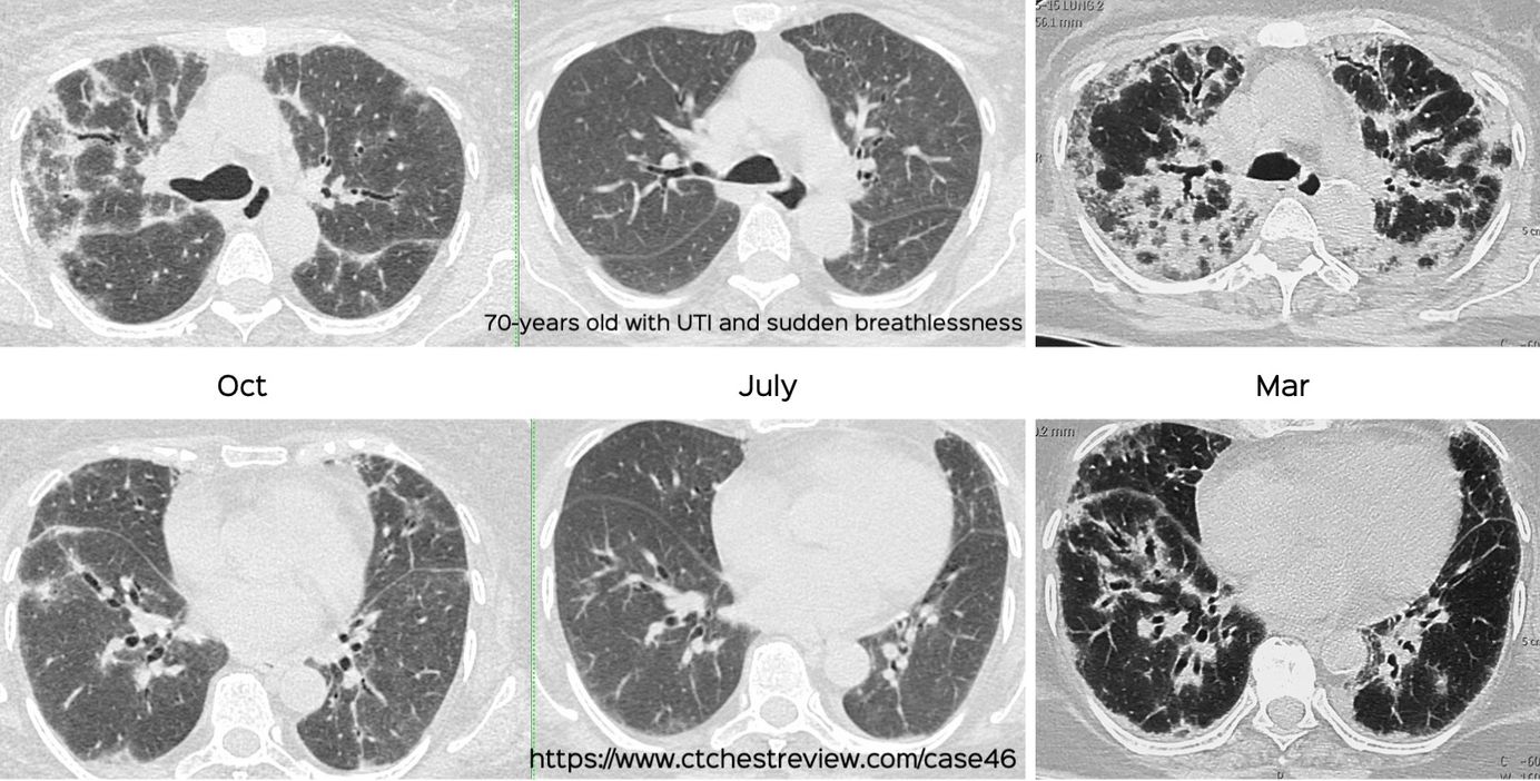
Case 46: The Discordance Between Surgeons and Physicians Paid Members Public
A good history helps us identify etiologies of reversible ILDs
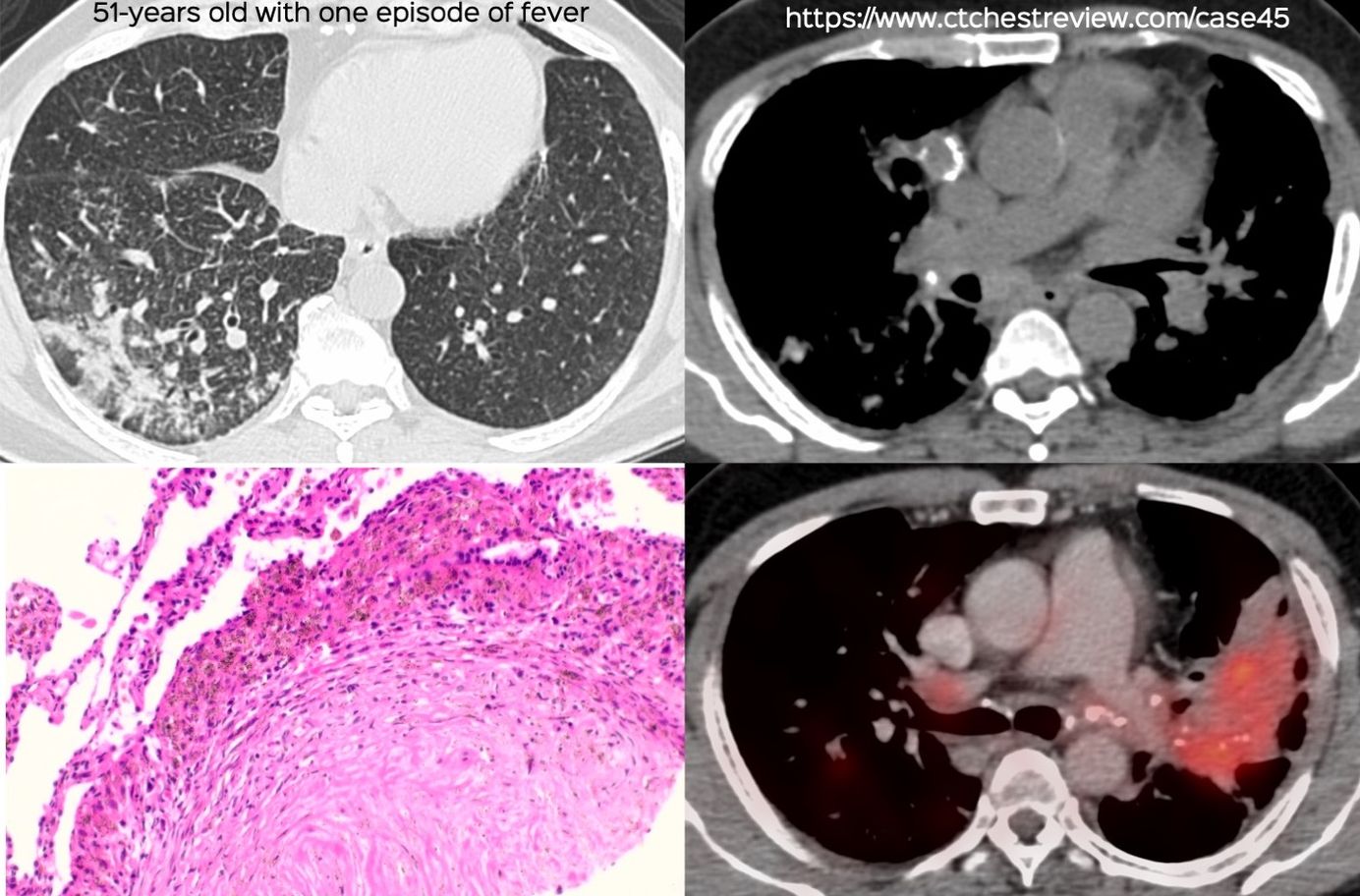
Case 45: The Power of Eggshell Calcification Paid Members Public
Recognizing eggshell calcification narrows the differential - it should trigger an occupational history search immediately.
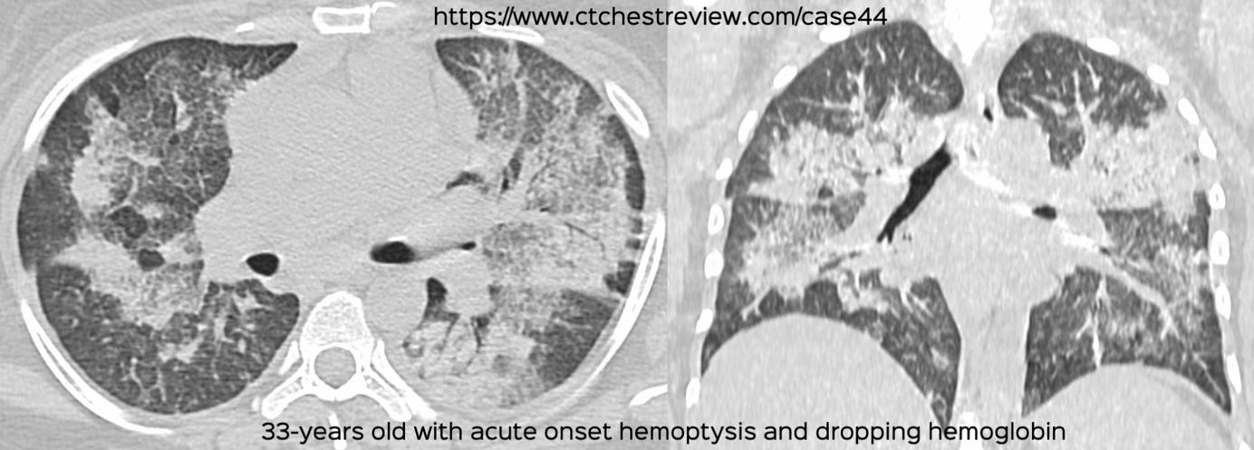
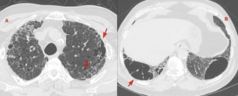
Case 44: Acute Onset Bilateral Ground Glass and Consolidation with Subpleural and Apical Sparing Paid Members Public
33-years old woman presented with sudden onset breathlessness.
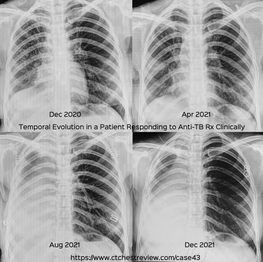
Case 43: Progressive Collapse in Patient Responding Clinically to Anti-Tuberculous Treatment Paid Members Public
Progressive Collapse in Patient Responding Clinically to Anti-Tuberculous Treatment
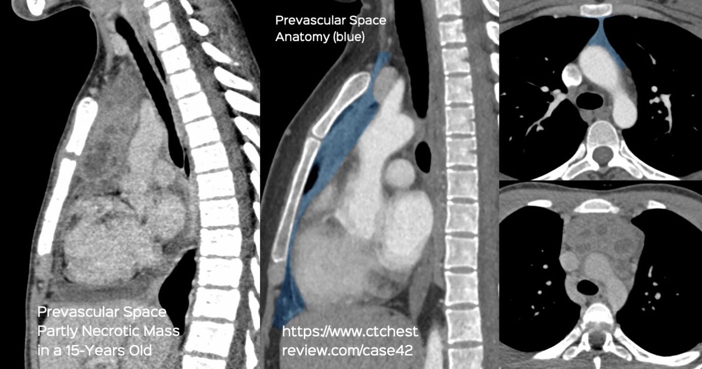
Case 42: Every Necrotic Mass in the Prevascular Space of the Mediastinum is NOT Tuberculosis Paid Members Public
Every Necrotic Mass in the Prevascular Space of the Mediastinum is NOT Tuberculosis
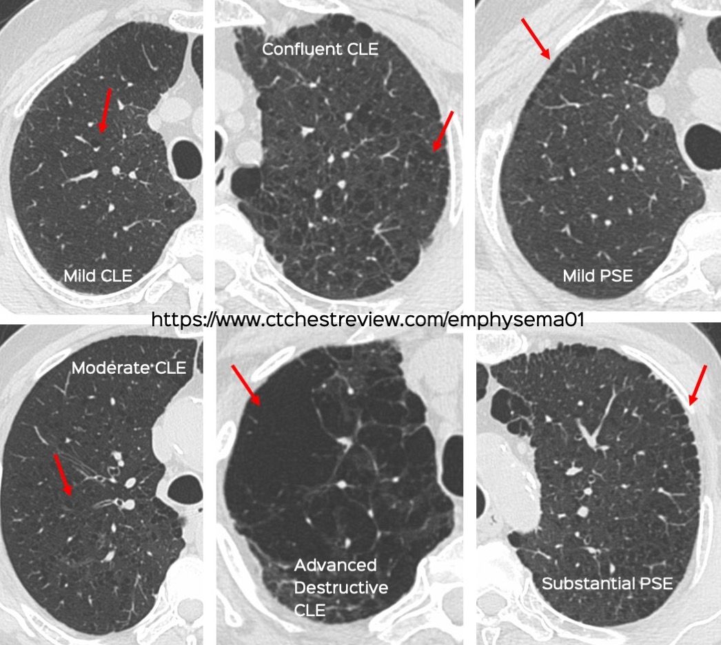
Snippet 12: Visual Classification of Centrilobular and Paraseptal Emphysema Paid Members Public
Visual Classification of Centrilobular and Paraseptal Emphysema
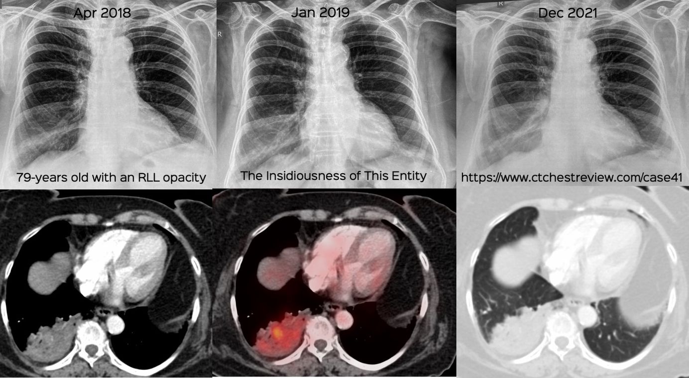
Case 41: The Insidiousness of this Entity Paid Members Public
Approach to a non-resolving opacitiy in the elderly
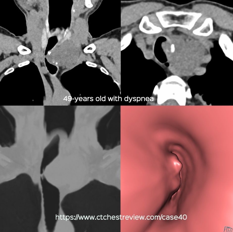
Case 40: Why is 2 + 2 Not Considered 4? Paid Members Public
A mediastinal mass where 2 + 2 = 4