Sarcoidosis - Two Cases - Stage II/III and Stage III/IV
The many appearances of sarcoidosis
Case 1:
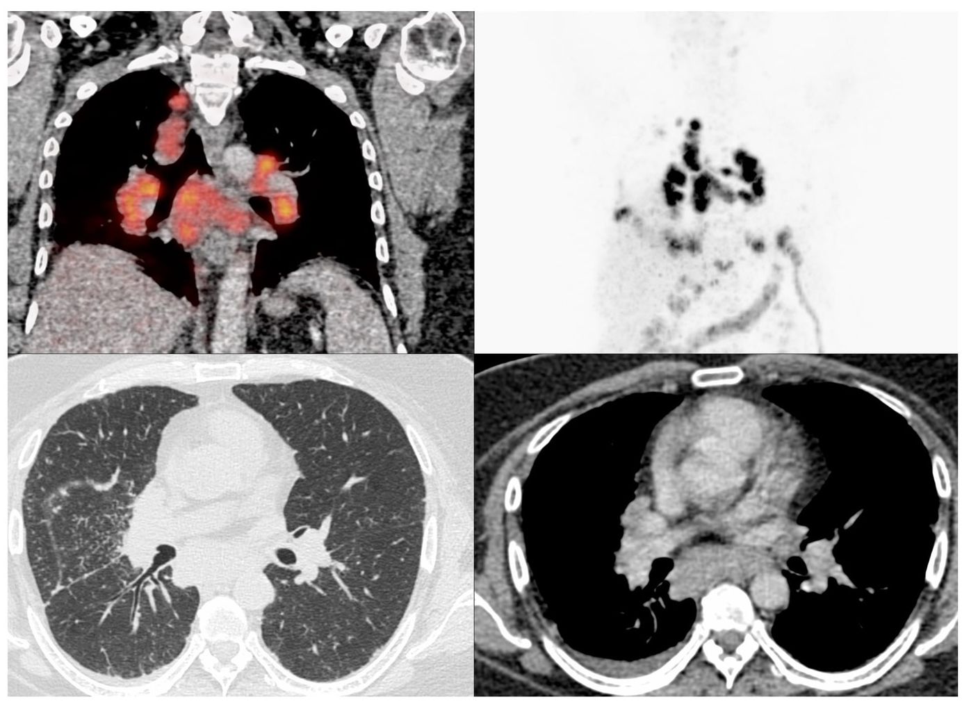
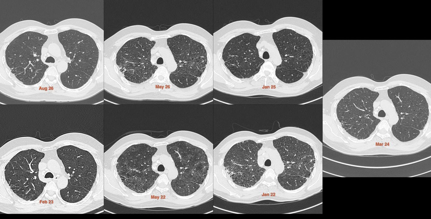
40-year-old in Jan 2022 presented with an unusual pattern of ground glass, fibrosis, nodules and cystic change. Known to have sarcoidosis since 2018. The nodes showed egg-shell calcification and were enlarged.
There was subtle improvement in the May 2022 scan of the lung findings with significant nodal improvement. The 2023 scan showed significant improvement, which remained stable in 2024 and 2025.
In May 2026, there was relapse, the picture looking similar to the Jan 2022 scan. The current Aug 2026 scan shows significant improvement again, the image resembling the 2023 scan.
The video is just a run-through the case on Osirix with all 7 scans shown together, scrolling back and forth to demonstrate the signs and the temporal evolution of the waxing, waning findings.
Case 2:
The next day was this case
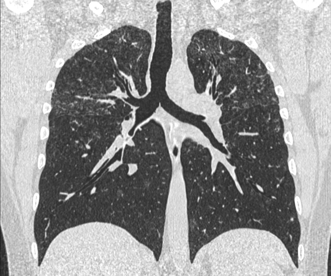
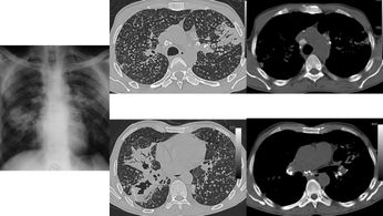
42-year-old in Jun 2022 presented a combination of upper lobe fissural and peribronchovascular nodules with fibrosis with basal fibrosis. He was known to have sarcoidosis and this would constitute Stage III/IV disease, though the Scadding classification does not accurately capture the situation, since he likely had both active disease and fibrosis without adenopathy.
The subsequent scans show gradual progression of both the upper lobe nodules and the lower lobe fibrosis.
The video is a run-through the case on Osirix with all 4 scans shown together, scrolling back and forth to demonstrate the signs and the temporal evolution and progression
This post is for subscribers only
SubscribeAlready have an account? Log in