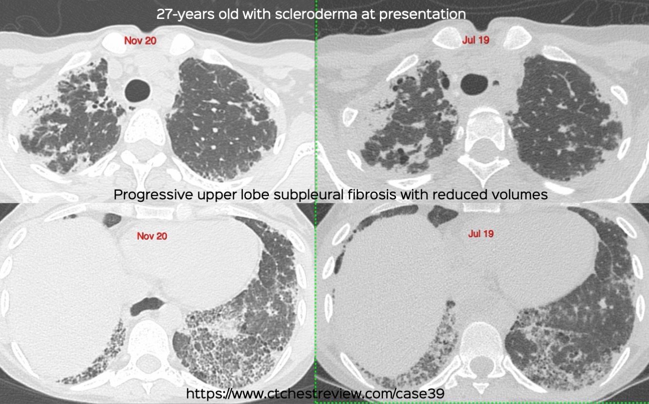
Case 39: Upper Lobe Subpleural Fibrosis Paid Members Public
27-years old with progressive upper lobe subpleural fibrosis
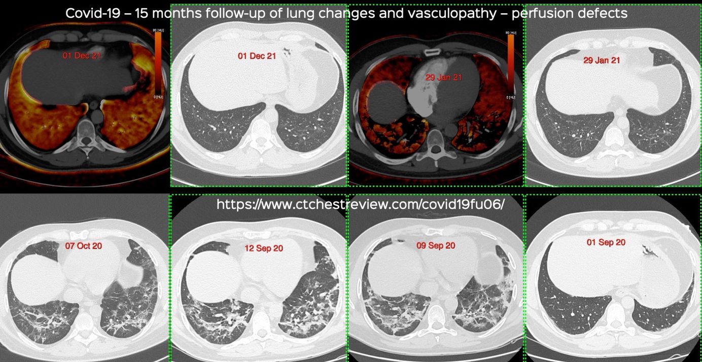
Case 38: Covid-19. 15 Months Follow-up of Lung Changes and Vasculopathy - Perfusion Defects Paid Members Public
15 months follow-up of lung changes and perfusion defects in a patient with Covid-19
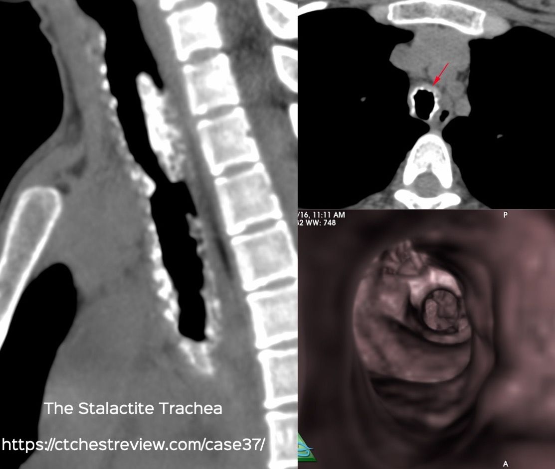
Case 37: The Stalactite Trachea Paid Members Public
The stalactite trachea
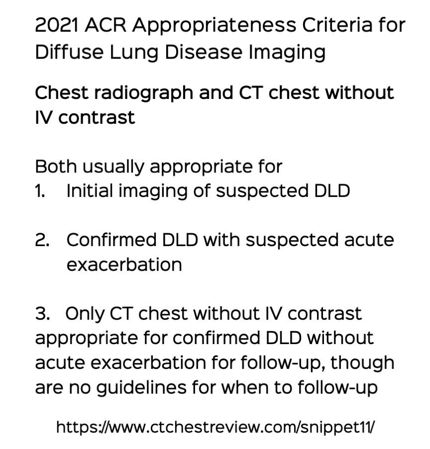
Snippet 11: ACR Appropriateness Criteria for Diffuse Lung Disease Imaging and ACR-STR Guidelines for Scanning Paid Members Public
CT chest without IV contrast is the primary modality for the evaluation of diffuse lung diseases and should be performed correctly to ensure proper interpretation
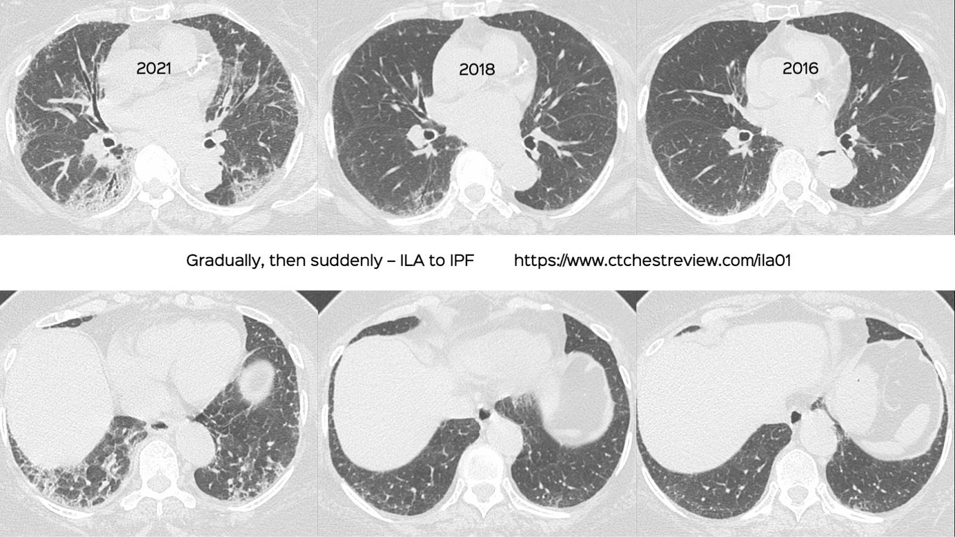
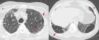
Case 36: Gradually, then Suddenly. ILA to ILD Paid Members Public
Gradually, then suddenly. ILA to ILD
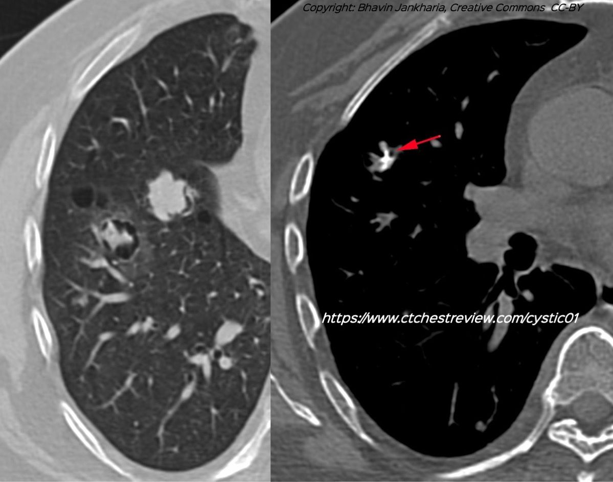
Case 35: Cysts and Nodules and Cysts with Nodules Paid Members Public
A patient with cysts and nodules and cysts with nodules
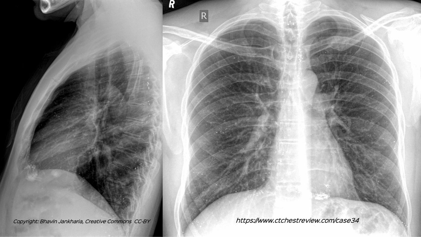
Case 34: All High-Density Foci in the Lungs Are Not Old Calcified Granulomas Paid Members Public
All high-density foci in the lungs are not old calcified granulomas
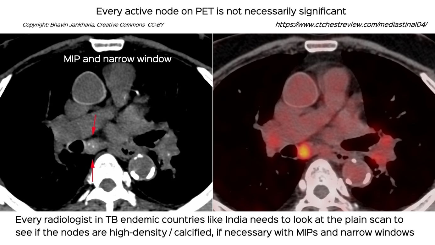
Case 33: Every Active Lymph Node on PET is Not Necessarily Significant Paid Members Public
Every active node on PET is not necessarily significant and needs to be evaluated for calcification / high density - features that suggest old healed disease